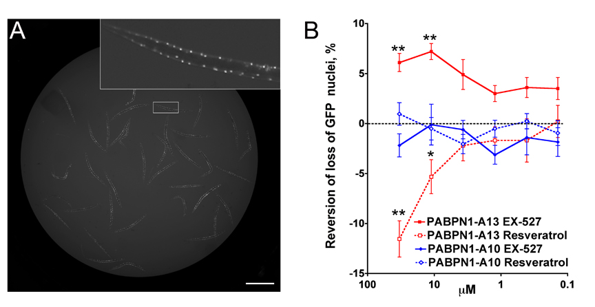
C. elegans is greatly suitable in suppressor screens and has strong potential to screen for the effects of modifiers at the subcellular level as it is transparent at all stages of lifespan. Here, we describe a 96-well plate assay that illustrates how high resolution imaging may be used in day-to-day research as well as RNAi and drug screens. We used the Plate Runner HD® (Trophos, France), a 96/384-well plate motorized device able to collect fluorescence at resolutions ranging from 1024x1024 px (1 px is 7.4 µm) to 8192x8192 px (1 px is 1 µm). This device has high depth of field (40 µm at resolution of 7.4 µm; 8 µm at resolution of 1 µm), thus allowing fluorescent signals to be quantified from whole animals. This device also has a wide-field objective that allows single images of the whole well to be acquired at once (Fig. 1A). To develop drug screening for neuromuscular disease, we used C. eleganstransgenics that co-express GFP and the oculopharyngeal muscular dystrophy (OPMD) protein PABPN1 in body wall muscle cells. Mutant PABPN1 animals show defective motility accompanied by loss of GFP nuclei (20 signals lost in 3-day adults) and muscle cell degeneration (Catoire et al., 2008). These phenotypes are aggravated by sirtuin (sir-2.1/SIRT1) activation and ameliorated by sirtuin inhibition (Catoire et al., 2008; Pasco et al., 2010). Resveratrol, an indirect SIRT1 activator, enhances mutant PABPN1 toxicity whereas EX-527, a selective SIRT1 inhibitor, has the opposite effect (Catoire et al., 2008; Pasco et al., 2010). We used these chemicals to normalize a drug screen assay (Fig. 1B). Synchronised L1 larvae are incubated at 20°C with bacteria and drugs (40 L1 in 50µl/well; 5 wells/point) until they reach adulthood. At 46 hours, FUDR (0.1 mg/ml) is added to prevent egg-laying and hatching. Day-3 adults are immobilized using 2,3-butanedione monoxime (0.1 M, 300 µl/well). Morphometric analysis of images (here 2048x2048) is performed using Image J and a script developed in the laboratory. Average numbers of GFP signals/worm are integrated into a database and subjected to statistical analysis. Drug screening is now ongoing using this assay. Perfect immobilization of animals is required for imaging at 4096x4096 and up. Similar strategies may be used in the development of a variety of suppressor screens.
Figures
References
Catoire H, Pasco MY, Abu-Baker A, Holbert S, Tourette C, Brais B, Rouleau GA, Parker JA, Néri C. (2008). Sirtuin inhibition protects from the polyalanine muscular dystrophy protein PABPN1. Hum. Mol. Genet. 17, 2108-17. 
Pasco MY, Rotili D, Altucci L, Farina F, Rouleau GA, Mai A, Néri C. (2010). Characterization of sirtuin inhibitors in nematodes expressing a muscular dystrophy protein reveals muscle cell and behavioral protection by specific sirtinol analogues. J. Med. Chem. 53, 1407-11.