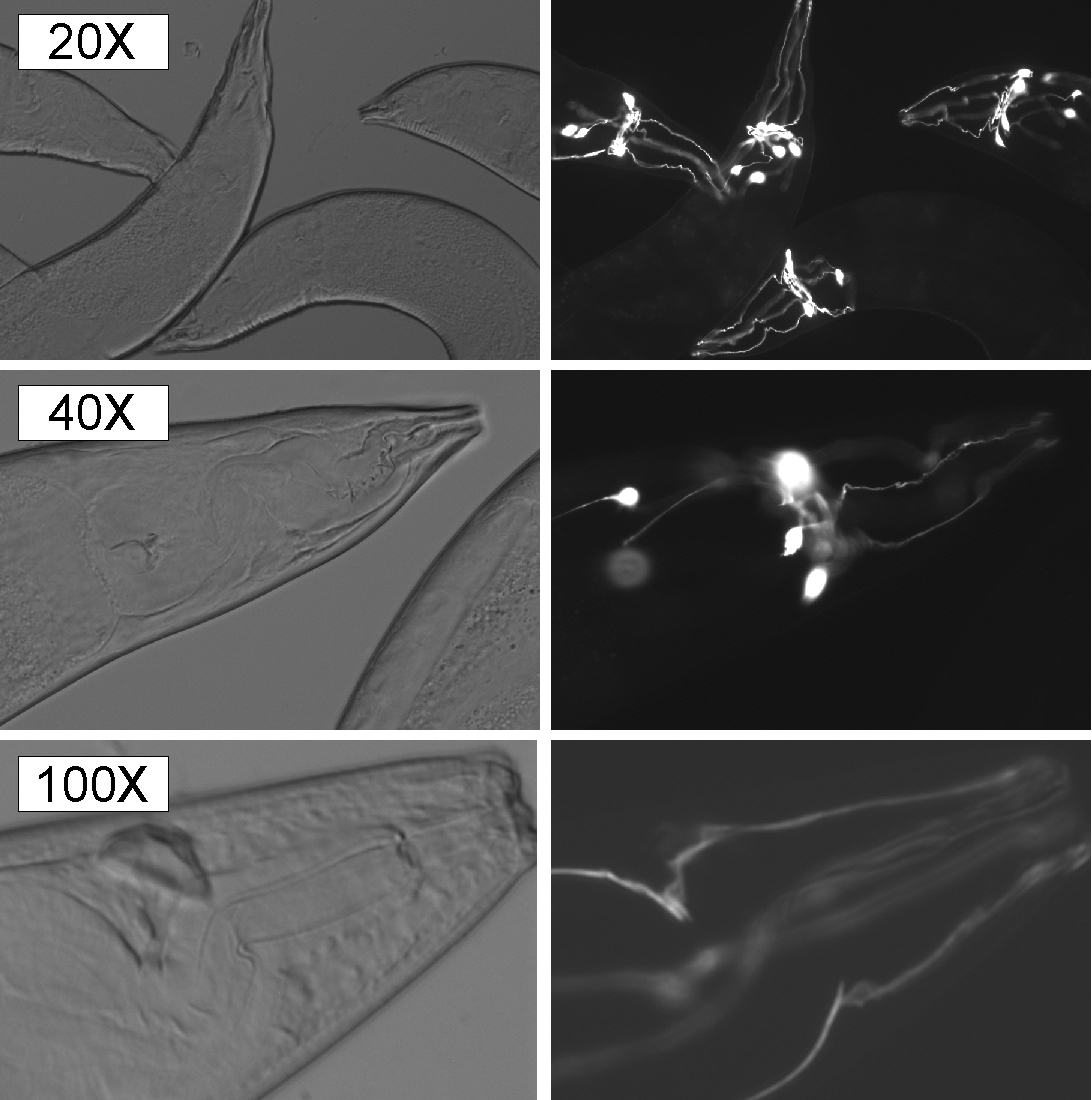
The use of 2% agarose pads is the conventional current method of choice for nematode microscopy. Although reliable, agarose pads are cumbersome to prepare and require the use of an immobilizing drug to keep nematodes motionless during the microscopy. Furthermore, a fast solidifying agarose drop on a glass slide requires some extra caution to make sure that the pad height remains minimum for microscopy with 40x or 100x lenses. We have developed a simpler and faster method for mounting nematodes for microscopy that does not require anesthetic drug. We report that a blend of equal percentage (20:20) of PEG 20000 and glycerol in 1x PBS provides enough viscosity to immobilize nematodes without distorting the cuticle/body shape. The treatment does not induce any observable osmotic shock when used on adult nematodes.
To perform the procedure, nematodes cleansed of bacteria are picked and then released into a PEG/glycerol droplet (6.5 μl) on a glass slide. A cover slip is then gently placed on the droplet containing the nematode. Due to the small volume of PEG/glycerol droplet, nematodes need to be transferred to PEG/glycerol droplet, and rapidly overlaid with a cover slip before mounting medium (PEG/glycerol) dries. This quick step instantly immobilizes the nematode and leaves them well suited for microcopy, including acquiring images for extended periods of time. We find the method is particularly useful for quick screening of GFP expressing lines, which frequently requires analysis of many nematodes, and use of high-resolution, compound microscopy for detecting low levels of GFP expression (such as due to certain weak promoters).
We have also experimented with other mixtures of PEG and glycerol. For instance, a mixture of 30% PEG 8000 in 25% glycerol also renders the nematodes amenable for microcopy, however increasing PEG concentrations above 30% renders nematodes unstable and unsuitable for microscopy. Larval nematodes appear to be slightly more sensitive to the procedure. We noticed slight cuticle distortion in younger nematodes (L1-L3) when PEG/Glycerol (20:20) solution was used for microscopy. A further dilution of PEG might be required to overcome the distortion and 1 μl of levamisole (20mM stock) can be used to insure that the nematodes are motionless.


