The soil environment of Caenorhabditis elegans in its native habitat is much more complex than either an agar or a liquid medium. Nematodes move within the three-dimensional soil space, responding not only to changes in medium (soil particles, water, colloidal mixtures), but also likely altering locomotory patterns in response to gravity, temperature, humidity, and other environmental cues. Notably, C. elegans accelerate away from blue and even shorter wavelengths of light, which are the wavelengths most likely to penetrate into soil (Edwards et al., 2008). The neural circuitry of the photophobic response is only just beginning to be studied. How other coincident information, like gravitational cues, might modulate locomotion is not known.
Most behavioral studies with C. elegans are performed using microscope-based imaging analysis, which are typically limited to a narrow vertical plane of focus. When observing worms in small droplets of liquid or in specially constructed channeled chambers, more locomotory behaviors arise than seen on flat surfaces (Lockery et al., 2008; Pierce-Shimomura et al., 2008). The worms navigate this more realistic environment with novel locomotory patterns, so-called “enhanced swimming” that relies on mechanosensory neuron activity (Park et al., 2008). However, the contribution or influence of the gravitational field and the movement of the worm within three-dimensional space cannot be studied easily with these microscope-based approaches (Kim et al., 2007).
How do worms behave when in a larger column of liquid, considerably larger than the dimensions of the worm? Do nematodes detect and respond to gravity? To study these kinds of questions, we have developed an inexpensive non-microscope-based imaging system that requires a laser, a cuvette, a lens and some mirrors, as well as a digital camera to record real-time images focused on a screen (Fig. 1). The full measurement technique is described in our two recent publications (Magnes et al., 2012b; Magnes et al., 2012a).
The diffraction-based measurements recorded the shape of the worms in space and time. We used shadow imaging, a modification of the diffraction-based method, to study the velocity of nematodes in a gravitational field. A small number of nematodes were placed in a quartz cuvette positioned in front of a laser of coherent light. The nematodes slowly fall down the column of water because of gravity, passing through the expanded laser beam. A digital camera records the moving patterns, which result from the locomotion of the worm. The velocities (speed and direction) are analyzed using Newtonian mechanics.
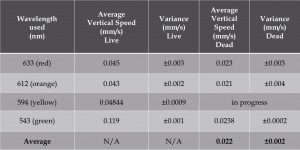
We assessed vertical velocity in wildtype worms using four different wavelengths of light and compared to velocities of worms killed with chloroform to control for passive effects of gravity on similar mass. We found that living nematodes locomote at twice the rate of passive fall in the column due to gravity alone. We also noted that velocities were greater when shorter wavelength lasers were used (Table 1).
Our preliminary findings indicate that worms are oriented “head down” towards the direction of the gravitational field. In contrast, dead worms, passively falling, fall in random orientations. These results suggest that nematodes actively move in the direction of gravity.
Figures

References
Edwards SL, Charlie NK, Milfort SC, Brown BS, Gravlin CN, Knecht, JE, Miller KG. (2008) A novel molecular solution for ultraviolet light detection in Caenorhabditis elegans. PLOS Biol 6, 1715-1729. 
Kim N, Dempsey DM, Kuan CJ, Zoval JV, O’Rourke E, Ruvkin G, Madou MJ, Sze JY. (2007) Gravity force transduced by the MEC-4/MEC-10 DEG/ENaC channel modulates DAF-16/FoxO activity in Caenorhabditis elegans. Genetics 177, 835-845. 
Lockery SR, Lawton KJ, Doll JC, Faumont S, Coulthard SM, Thiele TR, Chronis N, McCormick KE, Goodman MB, Pruitt BL. (2008) Artificial dirt: microfluidic substrates for nematode neurobiology and behavior. J Neurophysiol 99, 3136-3143. 
Magnes J, Susman K, Eells R. (2012) Quantitative locomotion study of freely swimming micro-organisms using laser diffraction. J Vis Exp 68, e4412. 
Park S, Hwang H, Nam SW, Martinez F, Austin RH, Ryu WS. (2008) Enhanced Caenorhabditis elegans locomotion in a structured microfluidic environment. PLOS One 3, e2550. 
Pierce-Shimomura JT, Chen BL, Mun JJ, Ho R, Sarkis R, McIntire SL. (2008) Genetic analysis of crawling and swimming locomotory patterns in C. elegans. Proc Natl Acad Sci U S A 105, 20982-20987. 


