Embryo series courtesy of Einhard Schierenberg
Table of Contents
Abstract
The role of neuropeptides in modulating behavior is slowly being elucidated. With the sequencing of the C. elegans genome, the extent of the neuropeptide genes in C. elegans can be determined. To date, 113 neuropeptide genes encoding over 250 distinct neuropeptides have been identified. Of these, 40 genes encode insulin-like peptides, 31 genes encode FMRFamide-related peptides, and 42 genes encode non-insulin, non-FMRFamide-related neuropeptides. As in other systems, C. elegans neuropeptides are derived from precursor molecules that must be post-translationally processed to yield the active peptides. These precursor molecules contain a single peptide, multiple copies of a single peptide, multiple distinct peptides, or any combination thereof. The neuropeptide genes are expressed extensively throughout the nervous system, including in sensory, motor, and interneurons. In addition, some of the genes are also expressed in non-neuronal tissues, such as the somatic gonad, intestine, and vulval hypodermis. To address the effects of neuropeptides on C. elegans behavior, animals in which the different neuropeptide genes are inactivated or overexpressed are being isolated. In a complementary approach the receptors to which the neuropeptides bind are also being identified and examined. Among the knockout animals analyzed thus far, defects in locomotion, dauer formation, egg laying, ethanol response, and social behavior have been reported. These data suggest that neuropeptides have a modulatory role in many, if not all, behaviors in C. elegans.
Neuropeptides are short sequences of amino acids that function either directly or indirectly to modulate synaptic activity. In addition, neuropeptides may also function as primary neurotransmitters. As in mammalian systems, the number of predicted neuropeptides in C. elegans is well over one hundred (Li et al., 1999; Pierce et al., 2001); however, most of the neuropeptides fall into two large families: the insulin-like peptides (Pierce et al., 2001; Li et al., 2003) and the FMRFamide (Phe-Met-Arg-Phe-NH2)-related peptides or FaRPs, which are referred to as FLPs in C. elegans (Li et al., 1998; Li, 2005). The remaining non-insulin, non-FLP peptides are classified as the neuropeptide-like proteins or NLPs. The NLPs are a diverse group of neuropeptides that have little similarity among each other (Nathoo et al., 2001). With a few striking exceptions, the functions of the different neuropeptides remain largely unknown in C. elegans. This overview serves to summarize the current state of the neuropeptide field.
Neuropeptides are typically derived from larger precursor molecules, which undergo posttranslational processing and sometimes modifications to yield mature peptides (see Figure 1). A single neuropeptide precursor molecule can give rise to a single neuropeptide, multiple distinct neuropeptides, multiple copies of a single neuropeptide, or any combination thereof. As an additional mechanism to increase neuropeptide complexity in mammals, a single precursor molecule can be differentially cleaved to yield different sets of peptides in different cell types (Strand, 1999; Salio et al., 2006). Furthermore, in the mollusk Aplysia peptides from a single precursor molecule can be sorted into different nerve terminals (Sossin et al., 1990). Whether such differential processing and trafficking occurs in C. elegans is unclear.
Like mammalian precursor molecules (Steiner, 1998), the initial cleavages in C. elegans occur C-terminal to dibasic residues flanking the peptide sequence (Rosoff et al., 1993; Marks et al., 1995, 1997, 1998, 1999a, 2001; Husson et al., 2005, 2006); however, cleavages C-terminal to mono- and tribasic residues have also been reported (Rosoff et al., 1993; Marks et al., 1997, 2001). The enzymes responsible for the initial endoproteolytic cleavage are kex2/subtilisin-like proprotein convertases, which must themselves be cleaved to become active. Cleavage of proprotein convertase is dependent on a chaperonin protein SBT-1 7B2 (Lindberg et al., 1998; Sieburth et al., 2005). Four C. elegans proprotein convertases, kpc-1, egl-3/kpc-2, aex-5/kpc-3, and bli-4/kpc-4, are present in C. elegans (Thacker and Rose, 2000). egl-3/kpc-2 is expressed in many, but not all neurons in the nervous system (Kass et al., 2001). Loss of egl-3/kpc-2 results in defects in egg-laying, mechanosensation, and locomotion (Kass et al., 2001; Jacob and Kaplan, 2003), indicating that EGL-3/KPC-2 cleaves precursors whose peptides have diverse functions. Moreover, using a polyclonal anti-FMRFamide antibody that recognizes the Arg-Phe-amide but not the non-amidated Arg-Phe-OH moiety (Marder et al., 1987), Kass and co-workers (2001) found decreased FMRFamide-like immunoreactivity in egl-3/kpc-2 mutants, suggesting that egl-3/kpc-2 cleaves some, but not all FLP precursors and that other proprotein convertases are active in the same cells. Among the other proprotein convertases, a kpc-1 deletion mutant shows mild locomotory defects and slow growth, suggesting that KPC-1 cleaves precursors of peptides involved in movement and growth (Thacker and Rose, 2000). Mutations in aex-5/kpc-3 cause defecation defects (Thomas, 1990). aex-5/kpc-3 is expressed in muscle (Thacker and Rose, 2000), and has been proposed to cleave a precursor molecule in muscle to produce a peptide that serves as a retrograde signal to regulate exocytosis (Doi and Iwasaki, 2002). A major function of BLI-4/KPC-4 is to cleave procollagen into collagen so that it can be deposited in the cuticle to give the cuticle structural integrity. Hence, null alleles of bli-4/kpc-4 cause lethality (Thacker et al., 1995). However, transcripts of bli-4/kpc-4 are also expressed in the nervous system (Thacker et al., 1995; Thacker and Rose, 2000), although phenotypes associated with loss of bli-4/kpc-4 neural transcripts are unknown. Collectively, these data indicate that multiple proprotein convertases are active in neurons.
|
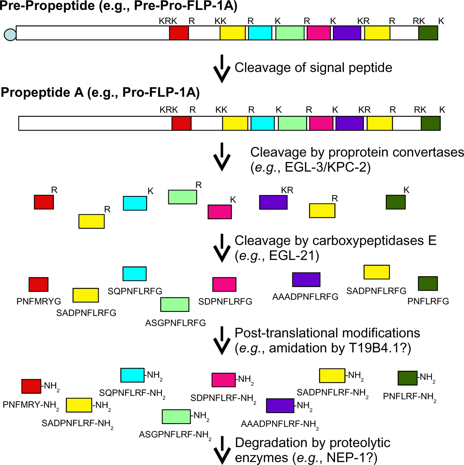
Figure 1. Processing of a neuropeptide gene product: flp-1 as an example. After translation of the flp-1A transcript, pre-pro-FLP-1A is cleaved by signal peptidase in the endoplasmic reticulum to release the signal peptide. The propeptide pro-FLP-1A is further cleaved C-terminal to mono-, di-, or tribasic residues (indicated by K and R) by proprotein convertases, such as EGL-3/KPC-2. The basic amino acids are removed by carboxypeptidases E, such as EGL-21, to yield the basic neuropeptides. The FLP-1A peptides are further modified by the addition of an amide group donated from the C-terminal glycine, a reaction which may be catalyzed by T19B4.1, to yield the active peptides. After release, peptides are removed from the synaptic cleft by proteolytic degradation, which may be mediated by NEP-1. With the exception of PNFMRFYamide, all flp-1A encoded peptides have been biochemically isolated (Li, 2005).
A peptidomic approach was also taken to determine the relative contribution of each proprotein convertase in neuropeptide processing (Husson et al., 2006). FLP and NLP peptides were isolated from wild type and different proprotein convertase mutants. kpc-1(gk8) and bli-4(e937)/kpc-4 mutants showed a similar peptide profile as wild type, suggesting that their contribution to peptide processing is minor or that the alleles examined, such as bli-4/kpc-4(e937), do not completely remove gene function. For two alleles of egl-3(n729 and gk238)/kpc-2 no peptides were isolated (Husson et al., 2006), confirming previous work that EGL-3/KPC-2 is the major active proprotein convertase in neurons (Kass et al., 2001). However, as previously reported (Kass et al., 2001), a residual level of FMRFamide-like immunoreactivity remained. Surprisingly, a much decreased peptide profile was seen in aex-5(sa23)/kpc-3 mutants (Husson et al., 2006), providing strong evidence that aex-5/kpc-3 is also responsible for neuropeptide precursor processing in neurons.
Once the precursor molecules are cleaved by the proprotein convertases, the basic residues themselves are removed from the peptide sequences by the activity of carboxypeptidase E. egl-21 encodes a neural-specific carboxypeptidase E that is expressed in about 60% of the neurons (Jacob and Kaplan, 2003). Loss of egl-21 carboxypeptidase E causes more severe phenotypes than those seen in the egl-3/kpc-2 proprotein convertase mutants. egl-21 null alleles show defects in egg laying, locomotion, mechanosensation, and defecation as well as extremely low levels of FMRFamide-like immunoreactivity (Jacob and Kaplan, 2003). These data suggest that cleavage by EGL-21, like EGL-3/KPC-2, yields peptides that function in multiple behaviors and that EGL-21 is one of the carboxypeptidases E that cleaves FLP precursor molecules. Two other carboxypeptidases are present in the C. elegans genome, but their roles in neuropeptide processing have not been investigated (Jacob and Kaplan, 2003).
To protect themselves from degradation, neuropeptides are commonly modified at the N- or C-terminus. In many circumstances, the modification also confers biological activity to the neuropeptide, including in C. elegans (Schinkmann and Li, 1992). The most common known modification in C. elegans is amidation. Based on the presence of a C-terminal glycine, which donates an amino group in the amidation process, all of the FLPs and many of the NLPs are likely to be amidated. In mammals two enzymes, peptidylglycine-alpha-hydroxylating monooxygenase (PHM) and peptidyl-alpha-hydroxyglycine alpha-amidating lyase (PAL) act sequentially to catalyze amidation; the enzymes are synthesized on the same molecule as adjacent domains on a bifunctional protein, peptidyl-α-hydroxyglycine α-amidating lyase (PAM; Eipper et al., 1993). C. elegans contains at least one PAM-like and one PHM molecule (Han et al., 2004). Decreased activity of T19B4.1, which encodes a monooxygenase, leads to resistance to the acetylcholinesterase inhibitor aldicarb, suggesting that T19B4.1 may be a PAM-like molecule that processes neuropeptides (Sieburth et al., 2005). Whether T19B4.1 and/or other enzymes are involved in neuropeptide amidation has not been determined.
Bioactive neuropeptides are located in dense core vesicles derived from the trans-Golgi network. By contrast, many of the classical small molecule transmitters are located in small, clear vesicles that are clustered at the synaptic zones. The processing of the neuropeptide precursor molecules starts in the endoplasmic reticulum with the removal of the signal peptide and continues in the Golgi complex and the dense core vesicles themselves as the vesicles are transported to the nerve terminal (Strand, 1999). UNC-104 kinesin is necessary for the transport of small, clear vesicles in C. elegans (Hall and Hedgecock, 1991) and can also function as the motor for dense core vesicles. Mutations in unc-104, for instance, cause an increase of FMRFamide-like immunoreactivity in neuronal cell bodies (Schinkmann, 1994; Jacob and Kaplan, 2003). Furthermore, fast, but not slow, anterograde transport of IDA-1, a transmembrane protein localized to dense core vesicles, is lost in unc-104 mutants, suggesting that at least two distinct motors can transport dense core vesicles (Zahn et al., 2004). Disruption of unc-116, which encodes a kinesin molecule distinct from UNC-104, decreases overall FMRFamide-like immunoreactivity (Schinkmann, 1994), suggesting that UNC-116 kinesin also plays a role in dense core vesicle trafficking.
In contrast to small clear vesicles, dense core vesicles are not localized at synaptic zones, but are more diffusely scattered around the nerve terminal (Strand, 1999; Salio et al., 2006). Whereas contents in small clear vesicles can be released by focal increases of calcium at the synaptic zone, release of neuropeptides from dense core vesicles appears to be dependent on a general increase of calcium throughout the nerve terminal, which can occur after high levels of stimulation (Strand, 1999; Salio et al., 2006). The exact mechanism for dense core vesicle movement to the cell membrane is unknown. Some mechanisms may be conserved for small clear and dense core vesicles. For instance, in C. elegans UNC-13 is necessary to prime both types of vesicles for release (Richmond et al., 1999; Sieburth et al., 2007). A cytoplasmic protein that promotes vesicle release by bridging between dense core vesicles and the plasma membrane is calcium-dependent activator protein (CAPS; Renden et al., 2001; Grishanin et al., 2002). Similarly, C. elegans UNC-31 CAPS also promotes dense core vesicle release (Sieburth et al., 2007); its activity appears to be modulated by IDA-1 (Cai et al., 2004). Mutations in pkc-1 protein kinase I cause increased punta fluorescence of neuropeptide precursor molecules, but not of GFP-SNB-1 puncta associated with synaptic vesicles, suggesting that PKC-1 is specifically necessary for dense core vesicle release (Sieburth et al., 2007). PKC-1 is expressed in the cholinergic ventral cord motor neurons and not the GABAergic motor neurons (Sieburth et al., 2007), indicating that other protein kinases function to promote dense core vesicle release in the GABAergic motor neurons. After release of the vesicle's contents, neuropeptides are cleared from the cleft by the action of proteolytic enzymes, one of which may be NEP-1 neprilysin (Sieburth et al., 2005). Hence, unlike small molecule transmitters, which can be recycled and re-loaded into synaptic vesicles, neuropeptides must be synthesized de novo in the cell body and transported down to the axon terminal.
In addition to their release at synapses, neuropeptides also act as hormones, i.e., as long range signaling molecules. Some of the first isolated mammalian neuropeptides, for instance, were hormones released from the pituitary, adrenal glands, and the gut (Strand, 1999). Similarly, neuropeptides in C. elegans released from neurons or non-neuronal cells (see below) may act as hormones (see below). To monitor expression of neuropeptides into the pseudocoelom, Sieburth et al. (2007) took advantage of the scavenger activity of the coelomocytes, which continuously endocytose fluid from the pseudocoelom (Fares and Grant, 2002), and monitored release of GFP-tagged neuropeptide precursors into the pseudocoelom by the appearance of GFP in the coelomocytes.
Before the sequencing of the C. elegans genome, one common method to identify neuropeptide candidates was to use different antibodies from the mammalian field to stain the C. elegans nervous system. Initial work reported immunoreactivity for cholecystokinin, Substance P, melanocyte-stimulating hormone, met-enkephalin, beta-endorphin, and possibly adrenocorticotopic hormone (S. McIntire, pers. comm.). Immunoreactivity was also detected for FMRFamide (Schinkmann and Li, 1992), which was initially isolated from invertebrates (Price and Greenberg, 1977) but for which related peptides were later found in mammals (Dockray, 2004). Subsequent sequencing of the C. elegans genome (C. elegans Sequencing Consortium, 1998) indicated that with the exception of Substance P- and FMRFamide-related peptides, none of these mammalian peptide families have been identified in C. elegans (Nathoo et al., 2001). Because of the similarity to the FLP neuropeptides, anti-cholecystokinin antibodies are likely to have cross-reacted with FLPs. The antigens to which the other mammalian antisera cross-reacted are unknown.
With the completion of the C. elegans genome, researchers were able to scan the genome for candidate genes encoding neuropeptides. Certain groups focused on specific neuropeptide families. For instance, several groups collectively identified forty genes that encode insulin-like molecules (see Table 1; Duret et al., 1998; Gregoire et al., 1998; Kawano et al., 2000; Pierce et al., 2001; Li et al., 2003). Our lab group identified twenty-four flp genes (flp-1 to flp-23 and flp-28) encoding peptides with a C-terminal RFamide moiety by cDNA isolation and BLAST searches (see Table 2; Li et al., 1998; Kim and Li, 2004; unpubl. obs.), while McVeigh and co-workers (2005; A. Maule, pers. comm.) used EST data mining to identify five additional flp genes, flp-24 to flp-27 and flp-32 (see Table 2). Husson and Schoofs (2007) identified flp-33 by isolating a FLP not encoded by any previously identified flp gene (see Table 2). Some of the flp genes also encode non-FLP peptides (see Table 2). Hart and co-workers used similarity and pattern-based scans in more general BLAST screens to identify other neuropeptide genes (Nathoo et al., 2001). Using the characteristics of neuropeptide precursor processing, the pattern-based scans were designed to search for peptide sequences that were flanked by mono- or dibasic sequences. 34 non-insulin-like, non-FLP-like genes were identified and are referred to collectively as the neuropeptide-like protein or nlp genes (Li et al., 1999; Nathoo et al., 2001; A. Hart, pers. comm.); an additional eight nlp genes were recently identified in a different screen and using peptidomic analysis (see Table 3; Couillault et al., 2004; Husson et al., 2005). A total of 113 neuropeptide genes encoding over 250 putative neuropeptides have now been identified in C. elegans (see Tables 1, 2 and 3).
What is striking about the neuropeptide genes is how many of them are clustered on a chromosome, suggesting that they arise from tandem gene duplications. In some cases, the neuropeptide genes are only a few hundred or thousand base pairs apart. For instance, within about 25,000 bp on chromosome I there are seven ins genes, ins-24 through ins-30, which do not appear to be part of an operon. Similarly, many of the nlp genes are also physically close (Nathoo et al., 2001). While the flp genes are clustered on certain chromosomes, particularly the X chromosome, only a few (e.g., flp-2, 3, and 28) are as physically close as some of the ins genes.
Because many of the flp neuropeptide genes encode multiple, distinct peptides, a nomenclature was developed to designate the distinct peptides produced by one gene. Each peptide is now designated by the gene name and a number. For instance, the flp-1 gene encodes eight distinct peptides designated as FLP-1-1, FLP-1-2, etc. (see Table 4). As more of the NLP and INS peptides are isolated and their sequences confirmed, these peptides may also be given specific designations.
Table 1. Neuropeptide genes encoding insulin-like peptides in C. elegans
|
Putative peptides | Expression Pattern | Function or Phenotype | Receptor | References## | |||||
|---|---|---|---|---|---|---|---|---|---|---|
|
|
ASI, ASJ, PQR, other neurons, hindgut, pharyngeal muscle, hypodermis | promotes repro-ductive growth | DAF-2 | 1 | |||||
|
|
ASI, ASJ, ASH, ADF, AIA, AIM, ASE, ASG, AWA, BAG, NSM, intestine, vulval muscles | DAF-2 antagonist? | DAF-2? | 2, 3, 4 | |||||
|
|
amphidial, labial, ventral cord, & tail neurons, pharynx, vulva | 2 | |||||||
|
|
amphidial, labial, lateral, ventral cord, & dorsal projecting neurons | 2 | |||||||
|
|
amphidial, labial, ventral cord, dorsal projecting, & tail neurons, hypodermis | DAF-2 | 2, 5 | ||||||
|
|
amphidial, labial, ventral cord, lateral projecting, & tail neurons, vulva | 2 | |||||||
|
|
amphidial, labial, ventral cord, & tail neurons | DAF-2 | 2 | ||||||
|
|
amphidial, labial, ventral cord, & tail neurons | 2 | |||||||
|
|
amphidial, labial, ventral cord, & tail neurons, vulva | 2 | |||||||
|
|
ASI, ASJ | Overexpression causes embryonic and larval arrest | DAF-2? | 2 | |||||
|
|
2 | ||||||||
|
|
labial, ventral cord, & tail neurons | 2 | |||||||
|
|
2 | ||||||||
|
|
2 | ||||||||
|
|
2 | ||||||||
|
|
2 | ||||||||
|
|
2 | ||||||||
|
|
2 | ||||||||
|
|
amphidial, ventral cord, tail, & pharyngeal neurons | DAF-2 antagonist? | DAF-2? | 2 | |||||
|
|
Overexpression causes larval arrest | DAF-2? | 2 | ||||||
|
|
2 | ||||||||
|
|
amphidial, ventral cord, & tail neurons | 2 | |||||||
|
|
amphidial, labial, ventral cord, lateral process projecting, & tail neurons | Modulates acetyl-choline signaling | 2, 6 | ||||||
|
|
amphidial, labial, & ventral cord neurons | 2 | |||||||
|
|
2 | ||||||||
|
|
2 | ||||||||
|
|
2 | ||||||||
|
|
2 | ||||||||
|
|
2 | ||||||||
|
|
2 | ||||||||
|
|
2 | ||||||||
|
|
Overexpression causes larval arrest$; Modulates acetyl-choline signaling | DAF-2? | 2$; 6 | ||||||
|
|
2$ | ||||||||
|
|
2$ | ||||||||
|
|
2, OST | ||||||||
|
|
2, OST | ||||||||
|
|
2 | ||||||||
|
|
2, OST | ||||||||
|
|
2 | ||||||||
|
|
2, OST | ||||||||
|
|
|||||||||
|
|
EST | ||||||||
| #Genes for which ESTs, ORFeomes (OST), or cDNAs have been isolated are in bold; encoded peptides are based on sequence homologies to other insulin-like peptides. Cosmid and LG data only indicated for ins-31 a; ins-31 b and ins-31 c are the same. $ Unclear which ins-31 construct was used for overexpression and functional data. See References. ##References are as follows: 1, Li et al., 2003; 2, Pierce et al., 2001; 3, Kodama et al., 2006; 4, Tomioka et al., 2006; 5, Kao et al., 2007; 6, Sieburth et al., 2005. EST or OST in EST or ORFeome databases indicated only if not identified in canonical reference and sequence spans at least one intron. Modified from Li (2005). | ||||||||||
Table 2. Neuropeptide genes encoding FMRFamide-related peptides (FLPs) in C. elegans
|
Putative peptides§ | Expression pattern† | Function or Phenotype | Receptor‡ | References## | ||||||||||||
|---|---|---|---|---|---|---|---|---|---|---|---|---|---|---|---|---|---|
|
|
AIA, AIY, AVA, AVE, AVK, RIG, RMG, M5 | involved in locomotion, egg laying, and fat deposition; SADPNFLRF-NH2 inhibits frequency of pharyngeal action potentials; modulates acetylcholine signaling | (C25G6.5, Y58G8A.4, C16D6.2, Y59H11AL.1) | 1-11, KA | ||||||||||||
|
|
AIA, RID, PVW, I5, MC (ASI, M4, head muscles, an extra pair of cells in the head) | T19F4.1a/b | 2, 9, 10, 12 | |||||||||||||
|
|
IL1, PQR; SP, CP9 | SAEPFGTMRF-NH2 inhibits frequency of pharyngeal action potentials | C53C7.1a (Y58G8A.4, C16D6.2) | 2,6-9, 12 | ||||||||||||
|
|
ADL, ASEL, AVM, AWC, FLP, PHA, PHB, PVD, I5, I6, NSM | C16D6.2 | 2,7,12 | |||||||||||||
|
|
PVT, RMG, I4, M4, pharyngeal muscle, amphidial neuron, (PB, I2); rays 1, 5, 7, HOB | GAKFIRF-NH2 increases frequency of pharyngeal action potentials | (C25G6.5) | 2,6-8,12 | ||||||||||||
|
|
ASE, AFD, ASG, PVT, I1 (one or two pairs of head cells); rays 2, 5, 6, 7 | increases frequency of pharyngeal action potentials | 2,6,8, 12,13 | |||||||||||||
|
|
ALA, AVG, PHB, PDA, PVW, RIC, SAA (RMDV/ SMDV, PHA) | Y59H11AL.1, C26F1.6 | 2, 9, 10, 12, 15 | |||||||||||||
|
|
AUA, PVM, URX (RMG/ADA, an extra pair of cells in the head); CP9 | increases frequency of pharyngeal action potentials; overexpression causes defecation defects | 2,6, 12, 16, AS, UP | |||||||||||||
|
|
inhibits frequency of pharyngeal action potentials; knockout shows slight sluggishness | (Y59H11AL.1) | 6, 8, 9, 12, 16, 17, UP | |||||||||||||
|
|
AIM, ASI, AUA, BAG, BDU, DVB, PQR, PVR, URX, vulD | 2,12 | ||||||||||||||
|
|
AUA, BAG, DA, DD, DVB, LUA, PHC, PVC, SAB, URX, VD, uv1, head muscle (socket cells); ray 4 | Y59H11AL.1, C26F1.6 (C16D6.2) | 2,6-9, 12, 15 | |||||||||||||
|
|
AVH/AVJ, BAG, PDA, PVR, SAA, SDQ, SMB (BDU); rays 1, 4, 5, 7, CP9 | 2, 12, 16 | ||||||||||||||
|
|
ASE, ASG, ASK, BAG, DD, I5, M3, M5 (an extra pair of cells in the head); VSP | APEASPFIRF-NH2 inhibits frequency of pharyngeal action potentials | (Y59H11AL.1) | 2, 6, 8, 9, 12, 16, 18, 19 | ||||||||||||
|
|
increases frequency of pharyngeal action potentials | (C25G6.5, C16D6.2) | 6, 7, 9, 16, 20, 21 | |||||||||||||
|
|
PHA, I2, socket/ sheath cells (pharyngeal muscle, several cells in the head) | C10C6.2 C16D6.2 | 2,7, 9, 20, 22 | |||||||||||||
|
|
AQTFVRF-NH2 inhibits frequency of pharyngeal action potentials | 6, 8, 20 | ||||||||||||||
|
|
BAG, M5 (an extra pair of cells in the head); rays 1, 5, 7 | 2, 20 | ||||||||||||||
|
|
AVA, AIY, RIG, RIM, M2 (M3, two extra pairs of cells in the head); rays 2, 6 | C16D6.2 Y58G8A.4 C53C7.1a NPR-1 (C25G6.5, F41E7.3) | 2, 6, 7, 9, 15, 19, 20, 23-26, UP | |||||||||||||
|
|
AIN, AWA, BAG, HSN, URX (an extra pair of cells in the tail); rays 5, 7, 9, CEM | 2, 6, 8, 9, 20 | ||||||||||||||
|
|
ALM, ASEL, AVM, LUA, PLM, PVC, PVM, PVR, RIB/AIB (PVT) | 2, 20, OH | ||||||||||||||
|
|
ADL, ASI, ASH, ASJ, ASK, FLP, URA, MC, M4, M2; CP6–9, SP, DVF | mutation causes mild aggregation behavior | NPR-1 C25G6.5 Y58G8a.4 | 2,7, 23-25 | ||||||||||||
|
|
AIM, ASG, AVA, AVG, AVL, CEP, PVD, PVW, RIC/AIZ, RIV, SMD, URA, uv1; 6 out of 9 CP | (Y59H11AL.1) | 2, 6, 8, 9, 20 | |||||||||||||
|
|
2, AM | |||||||||||||||
|
|
9, 27 | |||||||||||||||
|
|
9, 27 | |||||||||||||||
|
|
9, 27 | |||||||||||||||
|
|
9, 27 | |||||||||||||||
|
|
9, UP | |||||||||||||||
|
|
AM | |||||||||||||||
|
|
28 | |||||||||||||||
|
|
SH, LS | |||||||||||||||
| # Genes for which ESTs, ORFeomes (OST), cDNAs, or encoded peptides have been isolated are in bold. § Common sequences among peptides encoded by the same gene are indicated in blue. No. of copies of peptide encoded by gene indicated. A C-terminal glycine donates an amide group during amidation. † Based on co-localization with new markers, some expression patterns have been revised from published data (Kim and Li, 2004). Using an ASE-expressed red fluorescent protein, O. Hobert (pers. comm.) found that ASE did not express flp-5 or flp-21 and that only ASEL expressed flp-20. C. Bargmann (pers. comm.) confirmed that ASE did not express flp-21, and found that flp-21 was also expressed in ASK and ADL. Cells in parentheses are variably expressed and/or tentative identifications. Cells after semi-colons are male-specific. ‡ Receptors with an EC50≤1.5 μM are indicated; receptors with an EC50>1.5 μM are in parentheses. * Peptides have been biochemically isolated; *peptides including residues in parentheses have been isolated; non-FLP peptides are indicated in green. ## References are as follows: 1, Rosoff et al., 1992; 2, Kim and Li, 2004; 3, Rosoff et al., 1993; 4, Nelson et al., 1998b; 5, Waggoner et al., 2000; 6, Rogers et al., 2001; 7, Lowery et al., 2003; 8, Husson et al., 2005; 9, Husson et al., 2006; 10, Mertens et al., 2006; 11, Sieburth et al., 2005; 12, Nelson et al., 1998a; 13, Mertens et al., 2005; 14, Marks et al., 1998; 15, Mertens et al., 2004; 16, Davis and Stretton, 1996; 17, Marks et al., 1999a; 18, Marks et al., 1997; 19, Marks et al., 2001; 20, Li et al., 1999; 21, Marks et al., 1995; 22, Kubiak et al., 2003b; 23, Rogers et al., 2003; 24, Kubiak et al., 2003a; 25, de Bono and Bargmann, 1998; 26, Kubiak et al., 2008; 27, McVeigh et al., 2005; 28, Husson and Schoofs, 2007; pers. comm.: KA, K. Ashrafi; SH, Steven Husson; AM, A. Maule; LS, Lilianne Schoofs; UP, unpublished results; EST or OST in EST or ORFeome databases indicated only if not identified in canonical reference and sequence spans at least one intron. AF, Ascaris suum; PF, Panagrellus redivius. Modified from Li (2005). | |||||||||||||||||
Table 3. Neuropeptide Genes Encoding non-Insulin, non-FLP Peptides in C. elegans
|
Putative peptides§ | Expression pattern† | Function or Phenotype | References## | ||||||||||||
|---|---|---|---|---|---|---|---|---|---|---|---|---|---|---|---|---|
|
|
ASI, AWC, PHB, BDU, 4 head neurons, intestine | 1, 2, 2, 3 4 | |||||||||||||
|
|
1 head neuron, secretory cells near vulva, intestine | 2, OST | |||||||||||||
|
|
ADF, ASE, ASH, AWB, ASJ, BAG, HSN, I1, I2, I3, I4, MI, M3, NSMR, 3 head neurons, VNC, occ. I6, M2, pm1VL, intestine | 2 | |||||||||||||
|
|
2 | ||||||||||||||
|
|
ASI, 2 head neurons, spermatheca; 1 male tail neuron | 2 | |||||||||||||
|
|
ASI, IL1, 2 head neurons, 1 tail neuron, intestine | 1, 2, 3, 4 | |||||||||||||
|
|
ADL, AFD, ASE, ASI, PHA, VNC, 4 head neurons, 2 RVG neurons | 1, 2, 3, 4 | |||||||||||||
|
|
ASK, ADL, 6 head neurons, 2 tail neurons, I2, g1D, pm5L, pm5R, 2 RVG, processes in pharynx, intestine; HOB | 1, 2, 3, 4 | |||||||||||||
|
|
ASI, AWB, 4 head neurons, 1 tail neuron, VNC, spermatheca, vulval muscles, intestine | 2, 3, 4 | |||||||||||||
|
|
ASK, ADL, CAN, 2 lateral neurons, 1 tail neuron, 2 ant. pharyngeal neurons; 1 male tail neuron | 2 | |||||||||||||
|
|
IL1, 2 head neurons, VNC, PVD, 3 tail neurons, precomma embryos | 1, 2, 3, 4, EST, OST | |||||||||||||
|
|
1 tail neuron | Modulates acetylcholine signaling | 2, 4, 5 | ||||||||||||
|
|
3 head neurons, NSM, M2, I4, spermatheca, LUA, 1 tail cell, dorsal and ventral hypoderm, intestine | 2, 3, 4 | |||||||||||||
|
|
ASI, ASK, and another amphidial neuron, PHA, VNC, 2 RVG neurons, intestine | 1, 2, 3, EST | |||||||||||||
|
|
ASH, CAN, HSN, BDU, 5 head neurons, VNC, 3 RVG neurons, 1 tail neuron, intestine | 1, 2, 3 | |||||||||||||
|
|
7 head neurons, 1 lateral neuron, intestine | 2, 4 | |||||||||||||
|
|
1, 2, 3 | ||||||||||||||
|
|
ASI, 4 head neurons, 2 tail neurons, spermatheca, NSM, 2 ant. pharyngeal neurons, rectal gland, intestine | 1, 2, 3, 4 | |||||||||||||
|
|
4 head neurons, VNC in males, NSM, 4 post. pharyngeal neurons, spermatheca | 2, EST, OST | |||||||||||||
|
|
4 head neurons, 4 tail neurons, spermatheca, intestine, 1 ant. pharyngeal neuron | 2, 4 | |||||||||||||
|
|
AFD, 5 head neurons, VNC, 1 ant. pharyngeal neuron, 1 tail neuron, embryo, intestine | 1, 2, 3, 4 | |||||||||||||
|
|
2 | ||||||||||||||
|
|
tail, dorsal and ventral hypoderm | 2 | |||||||||||||
|
|
ASI, spermatheca, 1 pharyngeal neuron | anti-microbial? | 2, 6 | ||||||||||||
|
|
anti-microbial? | 2, 4, 6 | |||||||||||||
|
|
hypoderm | 2, 4 | |||||||||||||
|
|
ASI, 3 head neurons, spermatheca, hypoderm, intestine | anti-microbial? | 2, 6 | ||||||||||||
|
|
anti-microbial? | 2, 6, OST | |||||||||||||
|
|
hypoderm, intestine | anti-microbial? | 2, 6 | ||||||||||||
|
|
hypoderm | anti-microbial? | 2, 6 | ||||||||||||
|
|
hypoderm, embryos | anti-microbial | 2, 6 | ||||||||||||
|
|
anti-microbial? | 2, 6 | |||||||||||||
|
|
hypoderm | anti-microbial? | 6, EST, OST | ||||||||||||
|
|
EST,AH | ||||||||||||||
|
|
1, 3, 4 | ||||||||||||||
|
|
1, 3 | ||||||||||||||
|
|
1, 3, 4 | ||||||||||||||
|
|
1, 3, 4, AH, EST | ||||||||||||||
|
|
1, 3 | ||||||||||||||
|
|
1, 3, 4 | ||||||||||||||
|
|
1, 3, 4 | ||||||||||||||
|
|
AH, EST | ||||||||||||||
| # Genes for which ESTs, ORFeomes (OST), cDNAs, or encoded peptides have been isolated are in bold. § Common sequences among peptides encoded by the same gene are indicated in blue. No. of copies of peptide encoded by gene indicated. A C-terminal glycine donates an amide group during amidation. Some nlp peptide predictions have been revised. † Based on co-localization with new markers, some expression patterns have been revised from published data. Cells in parentheses are variably expressed and/or tentative identifications. * Peptides have been biochemically isolated; *peptides including residues in parentheses have been isolated; non-FLP peptides are indicated in green. ## References are as follows: 1, Li et al., 1999; 2, Nathoo et al., 2001; 3, Husson et al., 2005; 4, Husson et al., 2006; 5, Sieburth et al., 2005; 6, Couillault et al., 2004; pers. comm.: AH, Anne Hart; EST or OST in EST or ORFeome databases indicated only if not identified in canonical reference and sequence spans at least one intron. Modified from Li (2005). | ||||||||||||||||
Table 4. Peptides encoded by flp neuropeptide genes
|
Putative peptides§ | Peptide name | Name in other species | References## | |||||||||||||||||||||||||
|---|---|---|---|---|---|---|---|---|---|---|---|---|---|---|---|---|---|---|---|---|---|---|---|---|---|---|---|---|---|
|
|
|
|
1-6 | |||||||||||||||||||||||||
|
|
|
4, 7, 8 | ||||||||||||||||||||||||||
|
|
|
3, 4, 7, 8 | ||||||||||||||||||||||||||
|
|
|
7, 8 | ||||||||||||||||||||||||||
|
|
|
2, 7, 8 | ||||||||||||||||||||||||||
|
|
|
AF8/PF3 | 3, 7-11 | |||||||||||||||||||||||||
|
|
|
4, 7, 8 | ||||||||||||||||||||||||||
|
|
|
AF1 | 7, 8, 12, 13 | |||||||||||||||||||||||||
|
|
|
3, 4, 8, 12, 14 | ||||||||||||||||||||||||||
|
|
|
7, 8 | ||||||||||||||||||||||||||
|
|
|
|
3, 4, 7, 8, 10 | |||||||||||||||||||||||||
|
|
|
|
7, 8, 10, 12 | |||||||||||||||||||||||||
|
|
|
3, 4, 7, 8, 12, 15, 16 | ||||||||||||||||||||||||||
|
|
|
|
4, 12, 17-20 | |||||||||||||||||||||||||
|
|
|
4, 7, 17 | ||||||||||||||||||||||||||
|
|
|
|
3, 10, 17 | |||||||||||||||||||||||||
|
|
|
7, 17 | ||||||||||||||||||||||||||
|
|
|
|
4, 7, 16, 17, 21 | |||||||||||||||||||||||||
|
|
|
3, 4, 7, 17 | ||||||||||||||||||||||||||
|
|
|
7, 17 | ||||||||||||||||||||||||||
|
|
|
|
7, 9, 17 | |||||||||||||||||||||||||
|
|
|
3, 4, 7, 17 | ||||||||||||||||||||||||||
|
|
|
7, AM | ||||||||||||||||||||||||||
|
|
|
4, 22, UP | ||||||||||||||||||||||||||
|
|
|
4 | ||||||||||||||||||||||||||
|
|
|
4, 22 | ||||||||||||||||||||||||||
|
|
|
4, 22 | ||||||||||||||||||||||||||
|
|
|
4, UP | ||||||||||||||||||||||||||
|
|
|
AM | ||||||||||||||||||||||||||
|
|
|
23 | ||||||||||||||||||||||||||
|
|
|
SH, LS | ||||||||||||||||||||||||||
| # Genes for which ESTs, ORFeomes (OST), cDNAs, or encoded peptides have been isolated are in bold. § Common sequences among peptides encoded by the same gene are indicated in blue. No. of copies of peptide encoded by gene indicated. A C-terminal glycine donates an amide group during amidation. * Peptides have been biochemically isolated; *peptides including residues in parentheses have been isolated; non-FLP peptides are indicated in green. 1FLP-14/AF2 has also been found in Haemonchus contortus and Panagrellus redivius. ## References are as follows: 1, Rosoff et al., 1992; 2, Rosoff et al., 1993; 3, Husson et al., 2005; 4, Husson et al., 2006; 5, Geary et al., 1992; 6, Yew et al., 2005; 7, Kim and Li, 2004; 8, Nelson et al., 1998a; 9, Cowden and Stretton, 1995; 10, Yew et al., 2005; 11, Maule et al., 1994a; 12, Davis and Stretton, 1996; 13, Cowden et al., 1989; 14, Marks et al., 1999a; 15, Marks et al., 1997; 16, Marks et al., 2001; 17, Li et al., 1999; 18, Marks et al., 1995; 19, Cowden and Stretton, 1993; 20, Maule et al., 1994b; 21, Edison et al., 1997; 22, McVeigh et al., 2005; 23, Husson et al., 2007; pers. Comm.: SH, Steven Husson; AM, A. Maule; LS, Lilianne Schoofs; UP unpublished results. Modified from Li (2005). | |||||||||||||||||||||||||||||
To determine whether a candidate neuropeptide gene is transcribed and to confirm the genomic organization of the gene, two basic strategies have been used. One is to isolate cDNAs using reverse transcription (RT)-polymerase chain reaction (PCR); the second is to scan the C. elegans EST or ORFeome databases. Through these approaches, several groups have determined that 37 of the 40 insulin-encoding genes (Gregoire et al., 1998; Kawano et al., 2000; Pierce et al., 2001; Li et al., 2003), 28 of the 31 flp genes (Rosoff et al., 1992; Nelson et al., 1998a; Kim and Li, 2004; McVeigh et al., 2005; I. Miskelly, N.J. Marks, and A. Maule, pers. comm.; unpublished results), and 39 of the 42 nlp genes (Nathoo et al., 2001; Couillault et al., 2004) are expressed. Based on these data, we predict that most, if not all, of the candidate neuropeptide genes are expressed. The neuropeptide genes are relatively small and often have only a few exons. For instance, the coding and intronic regions of sixteen flp genes are less than 1 kilobase (Kim and Li, 2004; McVeigh et al., 2005). Among the nlp genes, a cluster of six nlp genes (nlp-27, 28, 29, 30, 31, and 34) on chromosome V encode very similar transcripts and have coding and intronic regions that range from 190 to 344 bp (Nathoo et al., 2001). The small size of most neuropeptide genes and the small number of exons that comprise the genes contribute to the difficulty of identifying all neuropeptide genes.
Because many of the C. elegans peptides have similar structures (for instance, the insulin-like peptides share common A and B domains and the FLPs all share a common C-terminal Arg-Phe-NH2), antibodies are difficult to generate against specific neuropeptides. Most peptide antibodies cross-react with several peptides of similar amino acid sequences. For example, the anti-FMRFamide antibody (Marder et al., 1987) recognizes FLPs encoded by several flp genes (Schinkmann and Li, 1992; Kim and Li, 2004). To determine the expression pattern of different neuropeptide genes, most researchers have made gene fusions of the promoter region of the neuropeptide gene to the coding region of a reporter gene, of which green fluorescent protein (GFP) is the most commonly used. The constructs are microinjected to generate transgenic animals. This relatively simple method has been used to determine the expression pattern of 60 neuropeptide genes (see Tables 1, 2 and 3), including 15 insulin-like genes (Pierce et al., 2001; Li et al., 2003), 19 flp genes (Kim and Li, 2004), and 27 nlp genes (Nathoo et al., 2001). For only one gene thus far, flp-8, has the GFP expression pattern been confirmed by immunocytochemistry with a FLP-8-specific monoclonal antibody (Sithigorngul et al., 1991; Kim, 2003).
Although there are inherent caveats to these expression patterns, a number of conclusions can be drawn from the data. First, the expression of neuropeptides is widespread in C. elegans and includes expression in the nervous system as well as in non-neuronal tissue (see Tables 1, 2 and 3). For instance, over 160 neurons, which represent over half of the 302 neurons in the C. elegans nervous system, express one or more FLPs (Kim and Li, 2004). Similarly, expression of the ins (Pierce et al., 2001), daf-28 (Li et al., 2003), and nlp (Nathoo et al., 2001) genes is widespread. Second, there is considerable overlap in the expression patterns. Although each gene appears to be expressed in a distinct set of neurons, a single neuron can express multiple neuropeptide genes and show a considerable diversity of neuropeptide expression. For instance, the chemosensory neuron ASI expresses daf-28, ins-1 and 9, nlp-1, 5, 6, 9, 14, 18, 24, and 27, and flp-2, 10, and 21 (Nathoo et al., 2001; Pierce et al., 2001; Li et al., 2003; Kim and Li, 2004); while some of these expression patterns may be artifactual, nevertheless ASI has the potential to release a plethora of neuropeptides to modulate neuronal activity. Third, neuropeptide expression is not limited to the nervous system. Neuropeptides, including those predicted by the ins, daf-28, flp, and nlp genes, are predicted to be in non-neuronal tissues, including intestine, somatic gonad, muscle, and hypodermis (Nathoo et al., 2001; Pierce et al., 2001; Li et al., 2003; Kim and Li, 2004). Secretion of neuropeptides from these tissues is likely to have an endocrine role.
Initially, the expression patterns of different peptides were compared in C. elegans and other nematodes by immunocytochemistry. The Ascaris nervous system, for instance, showed an extensive distribution of FMRFamide-like immunoreactivity (Cowden et al., 1993), much more than what was seen in C. elegans (Schinkmann and Li, 1992); however, the extent of flp expression with the GFP reporters in C. elegans now matches that in Ascaris. More recently, in situ hybridization has been used to examine the gene-specific expression pattern of different neuropeptide genes in the parasitic nematode Globodera pallida. For the few Gp-flp genes examined, there were several differences in the neurons in which the genes were expressed in C. elegans (Kimber et al., 2002). For instance, flp-6 is not expressed in C. elegans tail neurons, whereas Gp-flp-6 is expressed in several tail neurons (Kimber et al., 2002). The significance of these differences is unknown, but could suggest that the function of the peptides diverged in the related nematodes.
To determine whether the predicted peptides are produced, a few groups have begun the biochemical isolation of the different peptides. Thus far, most of the biochemical isolations have focused on the FLPs. To date, 36 FLPs encoded by seventeen flp genes (flp-1, 3, 5, 6, 8, 9, 11, 12, 13, 14, 16, 18, 19, 22, 24, 26, and 33) have been isolated (Rosoff et al., 1993; Marks et al., 1995, 1997, 1998, 1999a, 2001; Davis and Stretton, 1996; Husson et al., 2005; S. Husson and L. Schoofs, pers. comm.; Table 2). Indeed, the identification of flp-33 was based on the biochemical isolation of the peptide. Two FLPs that are not encoded by any of the identified flp genes have also been isolated (Davis and Stretton, 1996; N. Marks and A. Stretton, pers. comm.), underscoring the difficulty of identifying small neuropeptide genes with BLAST searches. In the recent peptidomic analysis by Husson and coworkers (2005), twenty nine NLPs encoded by 19 nlp genes were isolated (see Table 3), suggesting that many of the NLPs are also likely to be produced in C. elegans. These data also indicate that 113 is likely to be an underestimate of the total number of neuropeptide genes.
Peptides identical to some of the predicted FLPs have been isolated from related nematodes (see Table 4), such as Ascaris suum (Cowden et al., 1989; Cowden and Stretton, 1993, 1995; Yew et all., 2005), Haemonchus contortus (Keating et al., 1995; Marks et al., 1999b), and Panagrellus redivius (Geary et al., 1992; Maule et al., 1994a,b, 1995). To date, most of the isolated peptides in related nematodes belong to the FLP family (Yew et al., 2005). In addition, the FLP-12 (AF24) and FLP-21 (AF9) peptides have been isolated from Ascaris suum (Cowden and Stretton, 1995; Yew et al., 2005), suggesting that these peptides are also produced in C. elegans. Similarly, several of the predicted NLPs are similar to peptides isolated from other invertebrates (Nathoo et al., 2001). These data suggest that many of the predicted neuropeptides are indeed produced and highlight the rich diversity of neuropeptides in C. elegans.
Based on the expression pattern of the peptides, the peptides are likely to participate in a multitude of behaviors, including dauer formation, locomotion, egg-laying, and mechano- and chemosensation. To determine the function of the different neuropeptides, the most common strategy has been to inactivate or overexpress specific neuropeptide genes. This strategy has certain drawbacks with large neuropeptide superfamilies because of the possible functional overlap among the family members. Several neuropeptides, for instance, may bind and activate the same receptor. Using RNAi to decrease neuropeptide activity has not been routinely performed because of the inefficiency of RNAi in neurons (Simmer et al., 2002). Nevertheless, recent experiments have shown that several of the neuropeptide genes have unique functions.
Newly hatched first larval stage (L1) animals sample their environment to assess its qualities for reproductive growth. In the absence of food, newly hatched L1 animals will arrest reproductive growth and remain in this arrested state until a food source becomes available (Johnson et al., 1984). The decision to enter reproductive growth after hatching is dependent on the activity of DAF-2, an insulin-like receptor, and ASNA-1, an ATPase that acts non-cell autonomously and regulates the insulin pathway (see below; Gems et al., 1998; Kao et al., 2007).
Under continued exposure to harsh environmental conditions, such as overcrowding, high temperatures, or a scarce food supply during L1 or L2, C. elegans will undergo an alternative life cycle, referred to as the dauer life cycle (Cassada and Russell, 1975). After L2, animals enter the dauer state rather than L3 and remain in the dauer state until conditions improve, whereupon they exit the dauer state and resume the lifecycle as L4 animals (Cassada and Russell, 1975). The decision to enter reproductive growth or dauer is determined by parallel signaling pathways: the insulin and transforming growth factor β (TGF β) pathways (Riddle and Albert, 1997). Loss of either pathway results in constitutive dauer formation, indicating that the pathways function independently. Many of the insulin and TGFβ pathway mutants are temperature sensitive, and show an incompletely penetrant dauer phenotype at the permissive temperature (Riddle and Albert, 1997). Lowering the activity of an insulin and TGFβ pathway gene has a synergistic effect and causes a stronger dauer phenotype at the permissive temperature than lowering the activity of either gene alone (Thomas et al., 1993). Similarly, lowering the activity of asna-1 and a TGFβ pathway gene also leads to an enhanced dauer phenotype at the permissive temperature, thereby linking the L1 arrest and dauer decision to the insulin pathway (Kao et al., 2007). Hence, activation of DAF-2 leads to reproductive growth, whereas inactivation of DAF-2 leads to dauer arrest (Riddle and Albert, 1997). DAF-2 also functions to determine lifespan (Kenyon et al., 1993) and to limit body size (McCulloch and Gems, 2003). DAF-2 is the closest homologue to the mammalian insulin-like receptor superfamily (Kimura et al., 1997), but recently a more divergent family of 56 putative insulin-like receptors has been identified in C. elegans (Dlakic, 2002).
Members of the insulin superfamily are encoded by the ins genes and daf-28 in C. elegans; fifteen of the ins genes and daf-28 are expressed in neurons, including some of the amphidial chemosensory neurons (Pierce et al., 2001; Li et al., 2003). ins-1, ins-9, and daf-28 are expressed in ASI and ASJ chemosensory neurons, which are critical in the decision for dauer formation (Bargmann and Horvitz, 1991), as well as in other neurons; ins-1 and daf-28 are also expressed in non-neuronal tissue, such as intestinal cells (Pierce et al., 2001; Li et al., 2003). Only two insulin-family genes that have been inactivated by mutation have been examined thus far. INS-1 is most similar to mammalian insulin (Pierce et al., 2001). Loss of ins-1 has no effect on dauer formation or longevity (Pierce et al., 2001). By contrast, the daf-28(sa191) mutation causes transient dauer formation (Malone and Thomas, 1994). The mutation is likely to act as a dominant negative whereby the daf-28(sa191) gene product antagonizes DAF-2 activity (Li et al., 2003). Overexpression of daf-28 in a decreased TGFβ signaling background promotes exit from dauer, indicating that increased activity of the insulin pathway can bypass the TGFβ pathway (Kao et al., 2007). These data suggest that DAF-28 normally activates the DAF-2 insulin-like receptor to promote reproductive growth (Li et al., 2003). Consistent with this hypothesis, levels of a Pdaf-28::GFP transgene are decreased during starvation or application of dauer pheromone (Li et al., 2003), suggesting that daf-28 expression is regulated by environmental cues, as would be expected for a dauer regulator. Expression of the Pdaf-28::GFP transgene is found in an increasing number of cells as the animal ages (Li et al., 2003), suggesting that daf-28 is also involved in other behaviors, such as aging. ASNA-1 may regulate levels of DAF-28 in the pseudocoelom; asna-1 mutants show decreased levels of DAF-28::GFP in coelomocytes, indicating that less DAF-28::GFP is being released in the pseudocoelom (Kao et al., 2007).
If other insulin-like peptides also signal through the DAF-2 receptor, then perturbations of other ins genes may enhance or suppress the phenotypes of daf-2 and daf-28 mutants. The phenotypes caused by overexpression of several ins genes, using their endogenous promoters, were examined in different daf backgrounds. Overexpression of ins-1 and ins-18 caused a low level of dauer arrest and enhanced the dauer phenotype of daf-2 and/or daf-7 TGFβ mutants, whereas no dauer effects were seen with overexpression of ins-9, ins-19, ins-22, and ins-31 (Pierce et al., 2001). However, overexpression of ins-9 or of ins-31 and ins-19 in combination in a wild-type or daf-2 mutant background caused embryonic or larval arrest, a phenotype similar to one shown by some daf-2 alleles (Pierce et al., 2001). INS-1 and INS-18 may function to antagonize the activity of DAF-2 or to down-regulate daf-2 to promote dauer formation (Pierce et al., 2001), while INS-9, INS-31, and INS-19 may signal through DAF-2 to affect other aspects of development. Overexpression of ins-4 or ins-6 can suppress or partially suppress, respectively, the daf-28(sa191) mutation, suggesting that INS-4 or INS-6 can functionally substitute for DAF-28 and activate the DAF-2 receptor when present at high levels (Li et al., 2003). Furthermore, overexpression of ins-4 can bypass the effects of a mutation in the TGFβ pathway (Kao et al., 2007). By contrast, overexpression of ins-7, 9, 17, 21, 22, and 23 did not suppress the daf-28(sa191) mutation (Li et al., 2003). These data indicate that several insulin-like ligands signal through or affect DAF-2 activity to affect developmental growth and dauer formation, while other insulin-like ligands signal through other non-DAF-2 insulin receptors.
What are the roles of the other ins genes? Until mutants are isolated, the function of this large class of neuropeptides is largely unknown. Recently, INS-1 was identified as a key neuropeptide in the integration of behavior with the functional state of the animal. When placed on a thermal gradient well-fed animals move towards the temperature on which they were cultivated, whereas starved animals avoid the temperature at which they were cultivated (Hedgecock and Russell, 1975). This thermotaxis behavior is mediated by the thermosensory neuron AFD, which signals through the AIY interneuron (Mori and Ohshima, 1995). Well-fed ins-1 mutants exhibit normal thermotaxis, indicating that the basic thermosensory properties of AFD are intact in the mutants. Similar to starved wild-type animals, starved ins-1 animals slow when encountering food, demonstrating that ins-1 animals can recognize their starvation state (Kodama et al., 2006). However, starved ins-1 mutants move towards rather than away from their cultivation temperature, presumably because they cannot integrate cultivation temperature with starvation state (Kodama et al., 2006). This integration defect can be rescued by expression of ins-1 in different neurons and is partially suppressed by mutations in daf-2 and age-1, suggesting that INS-1 acts non-cell autonomously to antagonize signaling through the DAF-2 receptor (Kodama et al., 2006).
INS-1, however, can also activate the DAF-2 receptor. Wild-type animals normally chemotax towards sodium chloride (NaCl) (Ward, 1973). However, after pre-exposure to NaCl, starved, but not well-fed animals will avoid NaCl; this behavior is referred to as salt chemotaxis learning (Saeki et al., 2001). Several mutants, including daf-2, age-1, pdk-1, akt-1, and ins-1, are defective for salt chemotaxis learning (Tomioka et al., 2006), implicating involvement of the DAF-2 pathway in salt chemotaxis learning. Based on transgenic rescues and laser ablations, Tomioka and co-workers (2006) propose that INS-1 is released from AIA interneurons to activate DAF-2 receptors in ASER, thereby initiating the DAF-2 signaling cascade. Hence, INS-1 is involved in multiple integration events and whether it activates or antagonizes DAF-2 signaling is context dependent.
Acetylcholine is the primary excitatory transmitter at the neuromuscular junction in C. elegans. Aldicarb blocks the effects of acetylcholinesterase, thereby increasing the amount of acetylcholine at the synapse and causing paralysis and lethality (Nguyen et al., 1995). To identify genes that are resistant to aldicarb, a genome-wide RNAi screen was performed on eri-1;lin-15B or eri-1; dgk-1 lin-15B mutants, which have a sensitized background for RNAi (Sieburth et al., 2005). In addition to the processing enzymes, decreased activity of four neuropeptide genes, two ins genes (ins-22 and ins-31), one flp gene (flp-1), and one nlp gene (nlp-12), conferred aldicarb-resistance, suggesting that the peptides encoded by these genes modulate acetylcholine signaling (Sieburth et al., 2005).
Deletion mutants have been isolated for eleven flp genes (Nelson et al., 1998b; unpubl. obs.). Inactivation of flp-1 causes several defects, including hyperactive movement (Nelson et al., 1998b), defects in the timing of egg laying (Waggoner et al., 2000), thereby causing a decreased number of eggs laid (unpubl. obs.), and decreased fat stores in flp-1(yn2) mutants (K. Ashrafi, pers. comm.). FLP-1 peptides are also necessary for down-regulation of egg laying in the absence of food (Waggoner et al., 2000) and modulation of acetylcholine signaling (see above; Sieburth et al., 2005). The remaining flp mutants are currently being examined. Because so many flp genes have overlapping expression patterns, the function of these genes may also overlap and, therefore, be difficult to tease apart. Hence, deletion mutants are being screened on a large variety of behavioral assays. For instance, the swimming or thrashing assay, which involves placing animals in physiological buffer and counting the number of thrashes per minute, is more sensitive for detecting locomotion defects than examining the animal's movement on a solid surface. By the thrashing assay, flp-9 was found to thrash significantly less actively than wild-type animals (unpubl. obs.). Hence, to understand how the FLPs function, mutants will be examined for subtle defects, animals carrying multiple knockouts need to be isolated, and the receptor to which the peptides bind must be identified, as was done in the case of flp-21. The function of flp-21 will be discussed in conjunction with the function of its receptor, NPR-1.
Although no nlp mutant has been examined thus far, several of the nlp genes may function as anti-microbial peptides. In microarray analyses to identify genes whose expression levels are changed in response to fungal or bacterial insults, expression of nlp-29, 31, and 33 was induced (Couillault et al., 2004). Furthermore, the peptide encoded by nlp-31 has anti-microbial activity and protects against fungal infection (Couillault et al., 2004). The peptides encoded by nlp-24, 25, 27, 28, and 30 are similar to those encoded by nlp-29, 31, and 33, suggesting that these peptides also have anti-microbial functions (Couillault et al., 2004). nlp-29 is expressed in the hypoderm and intestine (Nathoo et al., 2001), nlp-31 in the hypoderm and embryos (Nathoo et al., 2001), and nlp-33 exclusively in the hypoderm (Couillault et al., 2004), suggesting that the peptides encoded by these genes only function as anti-microbial agents. By contrast, nlp-24 and 27 are also expressed in neurons (Nathoo et al., 2001) and may function both as anti-microbial agents and neuropeptides. The expression patterns of nlp-25 and 28 are unknown, so whether the peptides encoded by these genes also function as anti-microbial agents is unknown. As described above, nlp-12 is involved in modulating acetylcholine signaling (Sieburth et al., 2005).
As mentioned above, the function of a specific neuropeptide may be difficult to discern. Not only are multiple neuropeptides expressed in a single cell, but a specific neuropeptide may bind to multiple receptors. As illustrated above with the insulin-like peptides and their receptors, an alternative strategy to determine the function of neuropeptides is to inactivate the receptors to which the peptides bind.
Of the 1000 G-protein-coupled receptors in C. elegans, over 50 of them are likely to be neuropeptide receptors (Bargmann, 1998). Sixty G-protein receptors that were predicted to bind either a small molecule transmitter or a neuropeptide were inactivated by RNAi and screened for behavioral deficits (Keating et al., 2003). RNAi of six receptors, C16D6.2, C25G6.5, C26F1.6, F35G8.1, F41E7.3, and F59C12.2, resulted in either an increased or decreased brood size (Keating et al., 2003). Disruption by RNAi of eight receptors, AC7.1 (tachykinin-like), C15B12.5, C10C6.2, C24A8.4, F15A8.5, F59D12.1, T02E9.1, and T05A1.1, causes locomotion defects (Keating et al., 2003). The phenotypes from the RNAi data were confirmed in two cases by the isolation of deletion mutants in T05A1.1 and F35G8.1 (Keating et al., 2003). Several of the ligands for these receptors have now been identified (see below).
For the FLPs, many FaRP receptors have been isolated from other systems. With the exception of a molluscan FMRFamide-gated amiloride-sensitive channel that has homology to the MEC-4 and MEC-6 mechanoreceptors (Lingueglia et al., 1995), other identified FMRFamide receptors are G-protein coupled receptors (Tensen et al., 1998; Bonini et al., 2000; Cazzamali and Grimmelikhuijzen, 2002; Meeusen et al., 2002; Duttlinger et al., 2003). To match FLP ligands to specific G protein-coupled receptor binding partners, several groups have expressed candidate receptors in either heterologous cells or Xenopus oocytes (Kubiak et al., 2003a, b; Lowery et al., 2003; Rogers et al., 2003; Mertens et al., 2004; Table 2). Different FLP ligands were applied and several assays were used as the read out. Interestingly, all FLP receptors examined thus far can be activated by multiple FLPs. Some of the receptors are activated by multiple peptides encoded by one gene, while other receptors can be activated by peptides encoded by different genes. A few examples will be described below.
The Upjohn/Pharmacia group (Kubiak et al., 2003a, b; Lowery et al., 2003) transfected candidate receptors and chimeric G proteins into Chinese hamster ovary (CHO) cells, and used binding to GTPγS, a non-hydrolyzable form of GTP, as the readout. Binding of the cognate ligand to the receptor would presumably activate the receptor, thereby activating G proteins. GTPγS binding to membranes of transfected cells, therefore, indicates ligand binding. The group determined that peptides encoded by flp-15, GGPQGPLRFamide and RGPSGPLRFamide, activate C10C6.2 with an EC50 (concentration which produces 50% maximal activation) of 250 and 160 nM, respectively (Kubiak et al., 2003b). No other FLP activated the receptor, despite some FLPs, such as FLP-21 GLGPRPLRFamide, with high sequence similarity at the C-terminus (Kubiak et al., 2003b).
Mertens and coworkers (2004) expressed candidate receptors and Gα16 in HEK or CHO cells and screened for an increased calcium response, as monitored by an increase in fluorescence. The sensitivity of this system may be lower than that when using GTPγS binding as the readout, because the concentration of peptide needed to activate many receptors is generally much higher (usually in the μM rather than nM range). Nevertheless, Mertens and co-workers found that C26F1.6 binds to peptides encoded by two flp genes (Mertens et al., 2004). One flp-7-encoded peptide, FLP-7-2 TPMQRSSMVRFamide, activates C26F1.6 with an EC50 of ~1 μM; by contrast, FLP-7-1 SPMQRSSMVRFamide, which differs from FLP-7-2 by only one amino acid at the N-terminus, did not active the receptor at concentrations up to 10 μM. Interestingly, despite less sequence similarity, FLP-11-1 AMRNALVRFamide activates C26F1.6 with an EC50 of ~1.33 μM.
A FLP receptor can also bind to a diverse group of peptides with a range of activities. The Y59H11AL.1 receptor binds to 15 peptides encoded by 6 flp genes with EC50 values ranging from 25 nM to 5 μM (Mertens et al., 2006). Only three of the peptides, however, FLP-7-3 SPMERSAMVRFamide (25 nM), FLP-1-8 KPNFMRYamide (100 nM), and FLP-11-1 AMRNALVRFamide (750 nM) have EC50 values less than 1 μM (Mertens et al., 2006). While FLP-7-3 and FLP-11-1 have C-terminal sequence similarity, the structure of FLP-1-8 appears to be very different. Note that peptides encoded by flp-7 bind to two receptors, C26F1.6 and Y59H11AL.1. Where these two receptors are expressed may give us some clues as to how FLP-7 peptides affect behavior. Given that a FLP receptor can bind to multiple peptides encoded by different genes and a single peptide can bind to multiple receptors, the potential complexity of peptide actions in C. elegans is astounding.
de Bono and coworkers searched specifically for the ligand to the NPR-1 receptor, a homologue to the mammalian neuropeptide Y receptor. Mutations in the NPR-1 receptor affect aggregation behavior (de Bono and Bargmann, 1998) and tolerance to alcohol (Davies et al., 2004). On an agar plate with a bacterial food source, wild-type animals feed alone (referred to as solitary feeding); a mutation in npr-1 causes the animals to aggregate during feeding (referred to as social feeding) and accumulate at the edges of the bacteria (referred to as bordering behavior; de Bono and Bargmann, 1998). The aggregation behavior of npr-1 mutants can be suppressed by mutations in gcy-35 or gcy-36 (Cheung et al., 2004), both of which encode soluble guanylate cyclases (Morton et al., 1999). GCY-35 guanylate cyclase binds oxygen, and the aggregation behavior of npr-1 mutants may be related to oxygen levels in the local environment of the animals (Gray et al., 2004). To identify the NPR-1 ligand, the de Bono group injected constructs for NPR-1 as well as inwardly rectifying potassium channels into Xenopus oocytes, and screened for receptor activation of the potassium channels (Rogers et al., 2003). Because no neuropeptide Y is present in C. elegans, the assumption was that the NPR-1 ligand was a FLP. Both Rogers et al. (2003) and Kubiak et al. (2003a) determined that FLP-21 binds to NPR-1; in addition, Rogers et al. (2003) found that peptides encoded by flp-18 also activated NPR-1. Animals carrying a mutation in flp-21 display only mild aggregation compared to npr-1 mutants (Rogers et al., 2003). Two explanations may account for the difference in phenotypes between the FLP-21 ligand and the NPR-1 receptor mutants. The first is that the flp-21 mutation may be a partial loss of function allele instead of a null allele; the second is that FLP-18 ligands, which also bind NPR-1, may functionally substitute for the loss of FLP-21 ligands (Rogers et al., 2003). As with the other FLP receptors, NPR-1 is promiscuous in its binding to multiple FLP ligands produced by different flp genes. As with other FLPs, FLP-18 peptides bind multiple receptors, including NPR-1 (Kubiak et al., 2003a; Rogers et al., 2003) and Y58G8A.4 receptors (Kubiak et al., 2008).
Because many of the FLPs have been also found in parasitic nematodes (Cowden et al., 1989; Cowden and Stretton, 1993, 1995; Keating et al., 1995; Marks et al., 1999b) and, therefore, present possible targets for anti-helminthetic drugs, there has been an interest in examining the physiological effects of FLPs both in C. elegans as well as in parasitic nematodes. In C. elegans the effects of FLPs have been examined in a pharyngeal preparation (see Table 2; Rogers et al., 2001). Muscles of exposed pharyngeal terminal bulbs were impaled with microelectrodes to record changes of activity and action potential frequency. Surprisingly, many of the tested FLPs modulated the action potential frequency (Rogers et al., 2001). Peptides encoded by flp-5, 6, 8, and 14 increased the action potential frequency, while peptides encoded by flp-1, 3, 9, 13, and 16 decreased the action potential frequency. These data are consistent with the expression of some of these FLPs in the pharynx (Kim and Li, 2004) and suggest that multiple FLPs are involved in feeding behavior. Whether the different FLPs signal through one or multiple FLP receptors has yet to be determined.
Similarly, application of peptides encoded by 20 flp genes had a range of effects on body wall, reproductive, and pharyngeal muscle of Ascaris suum (Fellowes et al., 1998; Bowman et al., 2002; Moffett et al., 2003; Trailovic et al., 2005; for reviews see Maule et al., 1996; Brownlee et al., 1996; Brownlee and Walker, 1999; Brownlee et al., 2000), and it is likely that these FLPs have similar effects in C. elegans. On Ascaris muscle, different FLPs can activate different ion channels. Application of KPNFLRFamide (FLP-1-6) to somatic muscle cells, for instance, opens chloride channels, while application of SDPNFLRFamide (FLP-1-4) opens potassium channels (Bowman et al., 1996, 2002). The effects of KSAYMRFamide (FLP-6) on somatic muscle are context-dependent: application to ventral muscles causes contraction while application to dorsal muscles causes relaxation (Maule et al., 1994a). The response to FLPs can also be more complex. For instance, application of KNEFIRFamide (FLP-8) or KHEYLRFamide (FLP-14) to somatic muscle strips elicits biphasic responses consisting of an initial hyperpolarization, followed by an excitatory phase of rhythmic contractions (Cowden and Stretton, 1993; Bowman et al., 1996). More recently, Stretton and coworkers have begun to characterize the effects of C. elegans and Ascaris FLPs on the synaptic activity of Ascaris motor neurons (Davis and Stretton, 2001). Overall, the number of FLPs that can elicit physiological effects is striking and highlights the complex and intricate ways that different FLPs can modulate synaptic and muscle activity.
The diversity of neuropeptides in C. elegans rivals the numbers found in mammals. Although many of the peptides appear to be invertebrate specific, the insulin-like and FMRFamide-related peptides have counterparts in mammals. The daunting task of determining the regulation and functions of the different peptides remains. This task is complicated by the overlapping expression and redundant functions of the different peptides, as well as by their ability to bind multiple receptors, and, likewise, by the ability of the receptors to bind multiple ligands. While most, if not all, FLPs and NLPs are likely to signal through G protein-coupled receptors and the insulin-like ligands to signal through receptor tyrosine kinases, the identities of these receptors are still largely unknown. Despite the limited information available thus far, we envision that neuropeptides are involved in all behaviors in C. elegans.
We thank K. Ashrafi, A. Hart, A. Maule, P. McVeigh, S. McIntire, L. Schoofs, and A. Stretton for sharing unpublished data, O. Hobert with revisions of expression patterns, C. Ferguson for comments on the manuscript, and A. Hart and A. Maule for helpful discussions. This work was supported by grants from the NIH to C.L. and the City College of the City University of New York.
Bargmann, C.I. (1998). Neurobiology of the Caenorhabditis elegans genome. Science 282, 2028–2033. Abstract Article
Bargmann, C.I., and Horvitz, H.R. (1991). Control of larval development by chemosensory neurons in Caenorhabditis elegans. Science 251, 1243–1246. Abstract Article
Bonini, J.A., Jones, K.A., Adham, N., Forray, C., Artymyshyn, R., Durkin, M.M., Smith, K.E., Tamm, J.A., Boteju, L.W., Lakhlani, P.P., et al. (2000). Identification and characterization of two G protein-coupled receptors for neuropeptide FF. J. Biol. Chem. 275, 39324–39331. Abstract Article
Bowman, J.W., Friedman, A.R., Thompson, D.P., Maule, A.G., Alexander-Bowman, S.J., and Geary, T.G. (2002). Structure-activity relationships of an inhibitory nematode FMRFamide-related peptide, SDPNFLRFamide (PF1), on Ascaris suum muscle. Int. J. Parasitol. 32, 1765–1771. Abstract Article
Bowman, J.W., Friedman, A.R., Thompson, D.P., Ichhpurani, A.K., Kellman, M.F., Marks, N.J., Maule, A.G., and Geary, T.G. (1996). Structure-activity relationships of KNEFIRFamide (AF1), a nematode FMRFamide-related peptide, on Ascaris suum muscle. Peptides 17, 381–387. Abstract Article
Brownlee, D.J., Fairweather, I., Holden-Dye, L., and Walker, R.J. (1996). Nematode neuropeptides: Localization, isolation and functions. Parasitol. Today 12, 343–351. Abstract Article
Brownlee, D., Holden-Dye, L., and Walker, R. (2000). The range and biological activity of FMRFamide-related peptides and classical neurotransmitters in nematodes. Adv. Parasitol. 45, 109–80. Abstract
Brownlee, D.J., and Walker, R.J. (1999). Actions of nematode FMRFamide-related peptides on the pharyngeal muscle of the parasitic nematode, Ascaris suum. Ann. N. Y. Acad. Sci. 897, 228–238. Abstract Article
C. elegans Sequencing Consortium (1998). Genome sequence of the nematode C. elegans: A platform for investigating biology. Science 282, 2012–2018. Abstract Article
Cai, T., Fukushige, T., Notkins, A.L., and Krause, M. (2004). Insulinoma-associated protein IA-2, a vesicle transmembrane protein, genetically interacts with UNC-31/CAPS and affects neurosecretion in C. elegans. J. Neurosci. 24, 3115–3124. Abstract Article
Cassada, R.C., and Russell, R.L. (1975). The dauerlarva, a post-embryonic developmental variant of the nematode Caenorhabditis elegans. Dev. Biol. 46, 326–342. Abstract Article
Cazzamali, G, and Grimmelikhuijzen, C.J. (2002). Molecular cloning and functional expression of the first insect FMRFamide receptor. Proc. Natl. Acad. Sci. U.S.A. 99, 12073–12078. Abstract Article
Cheung, B.H.H., Arellano-Carbajal, F., Rybicki, I., and de Bono, M. (2004). Soluble guanylate cyclases act in neurons exposed to the body fluid to promote C. elegans aggregation behavior. Curr. Biol. 14, 1105–1111. Abstract Article
Couillault, C., Pujol, N., Reboul, J., Sabatier, L., Guichou, J.-F., Kohara, Y., and Ewbank, J.J. (2004). TLR-independent control of innate immunity in Caenorhabditis elegans by the TIR domain adaptor protein TIR-1, an ortholog of human SARM. Nat. Immunol. 5, 488–494. Abstract Article
Cowden, C., Sithigorngul, P., Brackley, P., Guastella, J., and Stretton, A.O. (1993). Localization and differential expression of FMRFamide-like immunoreactivity in the nematode Ascaris suum. J. Comp. Neurol. 333, 455–468. Abstract Article
Cowden, C., and Stretton, A.O. (1993). AF2, an Ascaris neuropeptide: isolation, sequence, and bioactivity. Peptides 14, 423–430. Abstract Article
Cowden, C., and Stretton, A.O. (1995). Eight novel FMRFamide-like neuropeptides isolated from the nematode Ascaris suum. Peptides 16, 491–500. Abstract Article
Cowden, C., Stretton, A.O., and Davis, R.E. (1989). AF1, a sequenced bioactive neuropeptide isolated from the nematode Ascaris suum. Neuron 2, 1465–1473. Abstract Article
Davies, A.G., Bettinger, J.C., Thiele, T.R., Judy, M.E., and McIntire, S.L. (2004). Natural variation in the npr-1 gene modifies ethanol responses of wild strains of C. elegans. Neuron 42, 731–743. Abstract Article
Davis, R.E., and Stretton, A.O. (1996). The motornervous system of Ascaris: eletrophysiology and anatomy of the neurons and their control by neuromodulators. Parasitology 113, S97–S117. Abstract
Davis, R.E., and Stretton, A.O. (2001). Structure-activity relationships of 18 endogenous neuropeptides on the motor nervous system of the nematode Ascaris suum. Peptides 22, 7–23. Abstract Article
de Bono, M., and Bargmann, C.I. (1998). Natural variation in a neuropeptide Y receptor homolog modifies social behavior and food response in C. elegans. Cell 94, 679–689. Abstract Article
Dlakic, M. (2002). A new family of putative insulin receptor-like proteins in C. elegans. Curr. Biol. 12, R155–R157. Abstract Article
Dockray, G.J. (2004). The expanding family of -RFamide peptides and their effects on feeding behaviour. Exp. Physiol. 89, 229–235. Abstract Article
Doi, M. and Iwasaki, K. (2002). Regulation of retrograde signaling at neuromuscular junctions by novel C2 domain protein AEX-1. Neuron 33, 249–259. Abstract Article
Duret, L., Guex, N., Peitsch, M.C., and Bairoch, A. (1998). New insulin-like proteins with atypical disulfide bond pattern characterized in Caenorhabditis elegans by comparative sequence analysis and homology modeling. Genome Res. 8, 348–353. Abstract
Duttlinger, A., Mispelon, M., and Nichols, R. (2003). The structure of the FMRFamide receptor and activity of the cardioexcitatory neuropeptide are conserved in mosquito. Neuropeptides 37, 120–126. Abstract Article
Edison, A.S., Messinger, L.A., and Stretton, A.O. (1997). afp-1: a gene encoding multiple transcripts of a new class of FMRFamide-like neuropeptides in the nematode Ascaris suum. Peptides 18, 929–935. Abstract Article
Eipper, B.A., Milgram, S.L., Husten, E.J., Yun, H.Y., and Mains, R.E. (1993). Peptidylglycine alpha-amidating monooxygenase: a multifunctional protein with catalytic, processing, and routing domains. Protein Sci. 2, 489–497. Abstract
Fares, H. and Grant, B. (2002). Deciphering endocytosis in Caenorhabditis elegans. Traffic 3, 11–19. Abstract Article
Fellowes, R.A., Maule, A.G., Marks, N.J., Geary, T.G., Thompson, D.P., Shaw, C., Halton, D.W. (1998). Modulation of the motility of the vagina vera of Ascaris suum in vitro by FMRF amide-related peptides. Parasitology 116, 277–287. Abstract Article
Geary, T.G., Price, D.A., Bowman, J.W., Winterrowd, C.A., Mackenzie, C.D., Garrison, R.D., Williams, J.F., and Friedman, A.R. (1992). Two FMRFamide-like peptides from the free-living nematode Panagrellus redivivus. Peptides 13, 209–214. Abstract Article
Gems, D., Sutton, A.J., Sundermeyer, M.L., Albert, P.S., King, K.V., Edgley, M.L., Larsen, P.L., and Riddle, D.L. (1998). Two pleiotropic classes of daf-2 mutation affect larval arrest, adult behavior, reproduction and longevity in Caenorhabditis elegans. Genetics 150, 129–155. Abstract
Gray, J.M., Karow, D.S., Lu, H., Chang, A.J., Chang, J.S., Ellis, R.E., Marietta, M.A., and Bargmann, C.I. (2004). Oxygen sensation and social feeding mediated by a C. elegans guanylate cyclase homologue. Nature 430, 317–322. Abstract Article
Gregoire, F.M., Chomiki, N., Kachinskas, D., and Warden, C.H. (1998). Cloning and developmental regulation of a novel member of the insulin-like gene family in Caenorhabditis elegans. Biochem. Biophys. Res. Commun. 249, 385–390. Abstract Article
Grishanin, R.N., Klenchin, V.A., Loyet, K.M., Kowalchyk, J.A., Ann, K., and Martin, T.F. (2002). Membrane association domains in Ca2+-dependent activator protein for secretion mediate plasma membrane and dense-core vesicle binding required for Ca2+-dependent exocytosis. J. Biol. Chem. 277, 22025–22034. Abstract Article
Hall, D.H., and Hedgecock, E.M. (1991). Kinesin-related gene unc-104 is required for axonal transport of synaptic vesicles in C. elegans. Cell 65, 837–847. Abstract Article
Han, M., Park, D., Vanderzalm, P.J., Mains, R.E., Eipper, B.A., and Taghert, P.H. (2004). Drosophila uses two distinct neuropeptide amidating enzymes, dPAL1 and dPAL2. J. Neurochem. 90, 129–141. Abstract Article
Hedgecock, E.M. and Russell, R.L. (1975). Normal and mutant thermotaxis in the nematode Caenorhabditis elegans. Proc. Natl. Acad. Sci. U.S.A. 72, 4061–4065. Abstract Article
Husson, S.J., Clynen, E., Baggerman, G., De Loof, A., and Schoofs, L. (2005). Discovering neuropeptides in Caenorhabditis elegans by two dimensional liquid chromatography and mass spectrometry. Biochm. Biophys. Res. Commun. 335, 76–86. Abstract Article
Husson, S.J., Clynen, E., Baggerman, G., Janssen, T., and Schoofs, L. (2006). Defective processing o fneuropeptide precursors in Caenorhabditis elegans lacking proprotein converase 2 (KPC-2/ELG-3): mutant analysis by mass spectrometry. J. Neurochem. 98, 1999–2012. Abstract Article
Husson, S.J., and Schoofs, L. (2007). Altered neuropeptide profile of Caenorhabditis elegans lacking the chaperone protein 7B2 as analyzed by mass spectrometry. FEBS Lett. 581, 4288–4292. Abstract Article
Jacob, T.C., and Kaplan, J.M. (2003). The EGL-21 carboxypeptidase E facilitates acetylcholine release at Caenorhabditis elegans neuromuscular junctions. J. Neurosci. 23, 2122–2130. Abstract
Johnson, T.E., Mitchell, D.H., Kline, S., Kernal, R., and Foy, J. (1984). Arresting development arrests aging in the nematode Caenorhabditis elegans. Mech. Ageing Dev. 28, 23–40. Abstract Article
Kao, G., Nordenson, C., Still, M., Rönnlund, A., Tuck, S., and Naredi, P. (2007). ASNA-1 positively regulates insulin secretion in C. elegans and mammalian cells. Cell 128, 577–587. Abstract Article
Kass, J., Jacob, T.C., Kim, P., and Kaplan, J.M. (2001). The EGL-3 proprotein convertase regulates mechanosensory responses of Caenorhabditis elegans. J. Neurosci. 21, 9265–9272. Abstract Article
Kawano, T., Ito, Y., Ishiguro, M., Takuwa, K., Nakajima, T., and Kimura, Y. (2000). Molecular cloning and characterization of a new insulin/IGF-like peptide of the nematode Caenorhabditis elegans. Biochem. Biophys. Res. Commun. 273, 431–436. Abstract Article
Keating, C.D., Holden-Dye, L., Thorndyke, M.C., Williams, R.G., Mallett, A., and Walker, R.J. (1995). The FMRFamide-like neuropeptide AF2 is present in the parasitic nematode Haemonchus contortus. Parasitology 111, 515–521. Abstract
Keating, C.D., Kriek, N., Daniels, M., Ashcroft, N.R., Hopper, N.A., Siney, El.J., Holden-Dye, L., and Burke, J.F. (2003). Whole-genome analysis of 60 G protein-coupled receptors in Caenorhabditis elegans by gene knockout with RNAi. Curr. Biol. 13, 1715–1720. Abstract Article
Kenyon, C., Chang, J., Gensch, E., Rudner, A., and Tabtiang, R. (1993). A C. elegans mutant that lives twice as long as wild type. Nature 366, 461–464. Abstract Article
Kim, K. (2003). Function of a FMRFamide-related neuropeptide gene family in Caenorhabditis elegans. Boston University thesis.
Kim, K., and Li, C. (2004). Expression and regulation of an FMRFamide-related neuropeptide gene family in Caenorhabditis elegans. J. Comp. Neurol. 475, 540–550. Abstract Article
Kimber, M.J., Fleming, C.C., Prior, Al, Jones, J.T., Halton, D.W., and Maule, A. (2002). Localisation of Globodera pallida FMRFamide-related peptide encoding genes using in situ hybridization. Int. J. Parasitol. 32, 1095–1105. Abstract Article
Kimura, K.D., Tissenbaum, H.A., Liu, Y., and Ruvkun, G. (1997). daf-2, an insulin receptor-like gene that regulates longevity and diapause in Caenorhabditis elegans. Science 277, 942–946. Abstract Article
Kodama, E., Kuhara, A., Mohri-Shiomi, A., Kimura, K.D., Okumura, M., Tomioka, M., Iino, Y., and Mori, I. (2007). Insulin-like signaling and the neural circuit for integrative behavior in C. elegans. Genes Dev. 20, 2955–2960. Abstract Article
Kubiak, T.M., Larsen, M.J., Nulf, S.C., Zantello, M.R., Burton, K.J., Bowman, J.W., Modric, T., and Lowery, D.E. (2003a). Differential activation of “social” and “solitary” variants of the Caenorhabditis elegans G protein-coupled receptor NPR-1 by its cognate ligand AF9. J. Biol. Chem. 278, 33724–33729. Abstract Article
Kubiak, T.M., Larsen, M.J., Zantello, M.R., Bowman, J.W., Nulf, S.C., and Lowery, D.E. (2003b). Functional annotation of the putative orphan Caenorhabditis elegans G-protein-coupled receptor C10C6.2 as a FLP15 peptide receptor. J. Biol. Chem. 278, 42115–42120. Abstract Article
Kubiak, T.M., Larsen, M.J., Bowman, J.W., Geary, T.G., and Lowery, D.E. (2008). FMRFamide-like peptides (FLPs) encoded on the flp-18 precursor gene activate two isoforms of the orphan Caenorhabditis elegans G-protein-coupled receptor Y58G8A.4 heterologously expressed in mammalian cells. Biopolymers, in press. Abstract Article
Li, C. (2005). The ever-expanding neuropeptide gene families in the nematode Caenorhabditis elegans. Parasitology 131 Suppl. S109–S127. Abstract Article
Li, W., Kennedy, S.G., and Ruvkun, G. (2003). daf-28 encodes a C. elegans insulin superfamily member that is regulated by environmental cues and acts in the DAF-2 signaling pathway. Genes Dev. 17, 844–858. Abstract Article
Li, C., Kim, K., Nelson, L.S. (1998). FMRFamide-related neuropeptide gene family in Caenorhabditis elegans. Brain Res. 848, 26–34. Abstract Article
Li, C., Nelson, L.S., Kim, K., Nathoo, A., and Hart, A.C. (1999). Neuropeptide gene families in the nematode Caenorhabditis elegans. Ann. N. Y. Acad. Sci. 897, 239–52. Abstract Article
Lindberg, I, Tu, B., Muller, L., and Dickerson, I.M. (1998). Cloning and functional analysis of C. elegans 7B2. DNA Cell Biol. 17, 727–734. Abstract
Lingueglia, E., Champigny, G., Lazdunski, M., and Barbry, P. (1995). Cloning of the amiloride-sensitive FMRFamide peptide-gated sodium channel. Nature 378, 730–733. Abstract Article
Lowery, D.E., Geary, T.G., Kubiak, T.M., and Larsen, M.J. (2003). Pharmacia & Upjohn Company, G protein-coupled receptor-like receptors and modulators thereof, United States, Patent 6,632,621.
Malone, E.A., and Thomas, J.H. (1994). A screen for nonconditional dauer-constitutive mutations in Caenorhabditis elegans. Genetics 136, 879–886. Abstract
Marder, E., Calabrese, R.L., Nusbaum, M.P., and Trimmer, B. (1987). Distribution and partial characterization of FMRFamide-like peptides in the stomatogastric nervous systems of the rock crab, Cancer borealis, and the spiny lobster, Panulirus interruptus. J. Comp. Neurol. 259, 150–163. Abstract Article
Marks, N.J., Maule, A.G., Geary, T.G., Thompson, D.P., Li, C., Halton, D.W., and Shaw, C. (1998). KSAYMRFamide (PF3/AF8) is present in the free-living nematode, Caenorhabditis elegans. Biochem. Biophys. Res. Commun. 248, 422–425. Abstract Article
Marks, N.J., Maule, A.G., Geary, T.G., Thompson, D.P., Davis, J.P., Halton, D.W., Verhaert, P., and Shaw, C. (1997). APEASPFIRFamide, a novel FMRFamide-related decapeptide from Caenorhabditis elegans: structure and myoactivity. Biochem. Biophys. Res. Commun. 231, 591–595. Abstract Article
Marks, N.J., Maule, A.G., Li, C., Nelson, L.S., Thompson, D.P., Alexander-Bowman, S., Geary, T.G., Halton, D.W., Verhaert, P., and Shaw, C. (1999a). Isolation, pharmacology and gene organization of KPSFVRFamide: a neuropeptide from Caenorhabditis elegans. Biochem. Biophys. Res. Commun. 254, 222–230. Abstract Article
Marks, N.J., Sangster, N.C., Maule, A.G., Halton, D.W., Thompson, D.P., Geary, T.G., and Shaw, C. (1999b). Structural characterisation and pharmacology of KHEYLRFamide (AF2) and KSAYMRFamide (PF3/AF8) from Haemonchus contortus. Mol. Biochem. Parasitol. 100, 185–194. Abstract Article
Marks, N.J., Shaw, C., Halton, D.W., Thompson, D.P., Geary, T.G., Li, C., and Maule, A.G. (2001). Isolation and preliminary biological assessment of AADGAPLIRFamide and SVPGVLRFamide from Caenorhabditis elegans. Biochem. Biophys. Res. Commun. 286, 1170–1176. Abstract Article
Marks, N.J., Shaw, C., Maule, A.G., Davis, J.P., Halton, D.W., Verhaert, P., Geary, T.G., and Thompson, D.P. (1995). Isolation of AF2 (KHEYLRFamide) from Caenorhabditis elegans: evidence for the presence of more than one FMRFamide-related peptide-encoding gene. Biochem. Biophys. Res. Commun. 217, 845–851. Abstract Article
Maule, A.G., Bowman, J.W., Thompson, D.P., Marks, N.J., Friedman, A.R., and Geary, T.G. (1996). FMRFamide-related peptides (FaRPs) in nematodes: occurrence and neuromuscular physiology. Parasitology 113, S119–S135. Abstract
Maule, A.G., Shaw, C., Bowman, J.W., Halton, D.W., Thompson, D.P., Geary, T.G., and Thim, L. (1994a). KSAYMRFamide: a novel FMRFamide-related heptapeptide from the free-living nematode, Panagrellus redivivus, which is myoactive in the parasitic nematode, Ascaris suum. Biochem. Biophys. Res. Commun. 200, 973–980. Abstract Article
Maule, A.G., Shaw, C., Bowman, J.W., Halton, D.W., Thompson, D.P., Geary, T.G., and Thim, L. (1994b). The FMRFamide-like neuropeptide AF2 (Ascaris suum) is present in the free-living nematode, Panagrellus redivivus (Nematoda, Rhabditida). Parasitology 109, 351–356. Abstract
Maule, A.G., Geary, T.G., Bowman, J.W., Marks, N.J., Blair, K.L., Halton, D.W., Shaw, C., and Thompson, D.P. (1995). Inhibitory effects of nematode FMRFamide-related peptides (FaRPs) on muscle strips from Ascaris suum. Invert. Neurosci. 1, 255–265. Abstract Article
McCulloch, D., and Gems, D. (2003). Body size, insulin/IGF signaling and aging in the nematode Caenorhabditis elegans. Exp. Gerontol. 38, 129–136. Abstract Article
McIntire, Sl.L., Jorgensen, E., Kaplan, J., and Horvitz, H.R. (1993). The GABAergic nervous system of Caenorhabditis elegans. Nature 364, 337–341. Abstract Article
McVeigh, P., Leech, S., Mair, G.R., Marks, N.J.,. Geary, T.G., and Maule, A.G. (2005). Analysis of FMRFamide-like peptide (FLP) diversity in phylum Nematoda. Int. J. Parasitol. 35, 1043–1060. Abstract Article
Meeusen, T., Mertens, I., Clynen, E., Baggerman, G., Nichols, R., Nachman, R.J., Huybrechts, R., De Loof, A., and Schoofs, L. (2002). Identification in Drosophila melanogaster of the invertebrate G protein-coupled FMRFamide receptor. Proc. Natl. Acad. Sci. U.S.A. 99, 15363–15368. Abstract Article
Mertens, I., Clinckspoor, I., Janssen, T., Nachman, R., and Schoofs, L. (2006). FMRFamide-related peptide ligands activate the Caenorhabditis elegans orphan GPCR Y59H11AL.1. Peptides 27, 1291–1296. Abstract Article
Mertens, I., Meeusen, T., Janssen, T., Nachman, R., and Schoofs, L. (2005). Molecular characterization of two G protein-coupled receptor splice variants as FLP2 receptors in Caenorhabditis elegans. Biochem. Biophys. Res. Commun. 330, 967–974. Abstract Article
Mertens, I., Vandingenen, A., Meeursen, T., Janssen, T., Luyten, W., Nachman, R.J., De Loof, A., and Schoofs, L. (2004). Functional characterization of the putative orphan neuropeptide G-protein coupled receptor C26F1.6 in Caenorhabditis elegans. FEBS Lett. 573, 55–60. Abstract Article
Moffett, C.L., Beckett, A.M., Mousley, A., Geary, T.G., Marks, N.J., Halton, D.W., Thompson, D.P., and Maule, A.G. (2003). The ovijector of Ascaris suum: multiple response types revealed by Caenorhabditis elegans FMRFamide-related peptides. Int. J. Parasitol. 33, 859–876. Abstract Article
Mori, I. and Ohshima, Y. (1995). Neural regulation of thermotaxis in Caenorhabditis elegans. Nature 376, 344–348. Abstract Article
Morton, D.B., Hudson, M.L., Waters, E. and O'Shea, M. (1999). Soluble guanylyl cyclases in Caenorhabditis elegans: NO is not the answer. Curr. Biol. 9, R546–R547. Abstract Article
Nathoo, A.N., Moeller, R.A., Westlund, B.A., and Hart, A.C. (2001). Identification of neuropeptide-like protein gene families in Caenorhabditis elegans and other species. Proc. Natl. Acad. Sci. U.S.A. 98, 14000–14005. Abstract Article
Nelson, L., Kim, K., Memmott, J., and Li, C. (1998a). FMRFamide-related gene family in the nematode Caenorhabditis elegans. Mol. Br. Res. 58, 103–111. Abstract Article
Nelson, L.S., Rosoff, M.L., and Li, C. (1998b). Disruption of a neuropeptide gene, flp-1, causes multiple behavioral defects in Caenorhabditis elegans. Science 281, 1686–1690. Abstract Article
Nguyen, M., Alfonso, A., Johnson, C.D., and Rand, J.B. (1995). Caenorhabditis elegans mutants resistant to inhibitors of acetylcholinesterase. Genetics 140, 527–535. Abstract
Pierce, S.B., Costa, M., Wisotzkey, R., Devadhar, S., Homburger, S.A., Buchman, A.R., Ferguson, K.C., Heller, J., Platt, D.M., Pasquinelli, A.A. et al. (2001). Regulation of DAF-2 receptor signaling by human insulin and ins-1, a member of the unusually large and diverse C. elegans insulin gene family. Genes Dev. 15, 672–678. Abstract Article
Price, D.A., and Greenberg, M.J. (1977). Structure of a molluscan cardioexcitatory neuropeptide. Science 197, 670–671. Abstract Article
Renden, R., Berwin, B., Davis, W., Kyoungsook, A., Chin, C.-T., Kreber, R., Ganetzky, B, Martin, T.F.J., and Broadie, K. (2001). Drosophila CAPS is an essential gene that regulates dense-cre vesicle release and synaptic vesicle fusion. Neuron 31, 421–437. Abstract Article
Richmond, J.E., Davis, W.S, and Jorgensen, E.M. (1999). UNC-13 I srequired for synaptic vesicle fusion in C. elegans. Nat. Neurosci. 2, 959–964. Abstract Article
Riddle, D.L., and Albert, P.S. (1997). Genetic and environmental regulation of dauer larva development. In C. elegans II, D.L. Riddle, T. Blumenthal, B.J. Meyer, and J.R. Priess, eds. (New York, NY: Cold Spring Harbor Laboratory Press), pp. 739–768.
Rogers, C.M., Franks, C.J., Walker, R.J., Burke, J.F., and Holden-Dye, L. (2001). Regulation of the pharynx of Caenorhabditis elegans by 5-HT, octopamine, and FMRFamide-like neuropeptides. J. Neurobiol. 15, 235–244. Abstract Article
Rogers, C., Reale, V., Kim, K., Chatwin, H., Li, C., Evans, P., and de Bono, M. (2003). Inhibition of Caenorhabditis elegans social feeding by FMRFamide-related peptide activation of NPR-1. Nat. Neurosci. 6, 1178–85. Abstract Article
Rosoff, M.L., Burglin, T.R., and Li, C. (1992). Alternatively spliced transcripts of the flp-1 gene encode distinct FMRFamide-like peptides in Caenorhabditis elegans. J. Neurosci. 12, 2356–2361. Abstract
Rosoff, M.L., Doble, K.E., Price, D.A., and Li. C. (1993). The flp-1 propeptide is processed into multiple, highly similar FMRFamide-like peptides in Caenorhabditis elegans. Peptides 14, 331–338. Abstract Article
Saeki, S., Yamamoto, M., and Iino, Y. (2001). Plasticity of chemotaxis revealed by paired presentation of a chemoattractant and starvation in the nematode Caenorhabditis elegans. J. Exp. Biol. 204, 1757–1764. Abstract
Salio, C., Lossi, L., Ferrini, F., and Merighi, A. (2006). Neuropeptides as synaptic transmitters. Cell Tissue Res. 326, 583–598. Abstract Article
Schinkmann, K. (1994). FMRFamide-like peptides in the nematodes Caenorhabditis elegans and Caenorhabditis vulgaris. Boston University, Boston, MA, Ph.D. thesis.
Schinkmann, K., and Li, C. (1992). Localization of FMRFamide-like peptides in Caenorhabditis elegans. J. Comp. Neurol. 316, 251–260. Abstract Article
Simmer, F., Tijsterman, M., Parrish, S., Koushika, S.P., Nonet, M.L., Fire, A., Ahringer, J., and Plasterk, R.H. (2002). Loss of the putative RNA-directed RNA polymerase RRF-3 makes C. elegans hypersensitive to RNAi. Curr. Biol. 12, 1317–1319. Abstract Article
Sieburth, D., Ch'ng, Q.L., Dybbs, M., Tavazoie, M., Kennedy, S., Want, D., Dupuy, D., Rual, J.-F., Hill, D.E., Vidal, M., Ruvkun, G., and Kaplan, J.M. (2005). Systematic analysis of genes required for synapse structure and function. Nature 436, 510–517. Abstract Article
Sieburth, D., Madison, J.M., and Kaplan, J.M. (2007). PKC-1 regulates secretion of neuropeptides. Nat. Neurosci. 10, 49–57. Abstract Article
Sithigorngul, P., Stretton, A.O., and Cowden, C. (1991). A versatile dot-ELISA method with femtomole sensitivity for detecting small peptides. J. Immunol. Methods 141, 23–32. Abstract Article
Sossin, W.S., Sweet-Cordero, A., and Scheller, R.H. (1990). Dale's hypothesis revisited: different neuropeptides derived from a common prohormone are targeted to different processes. Proc. Natl. Acad. Sci. U.S.A. 87, 4845–4848. Abstract Article
Steiner, D.F. (1998). The proprotein convertases. Curr. Opin. Chem. Biol. 2, 31–39. Abstract Article
Tensen, C.P., Cox, K.J., Smit, A.B., van der Schors, R.C., Meyerhof, W., Richter, D., Planta, R.J., Hermann, P.M., van Minnen, J., Geraerts, W.P., et al. (1998). The Lymnaea cardioexcitatory peptide (LyCEP) receptor: a G-protein-coupled receptor for a novel member of the RFamide neuropeptide family. J. Neurosci. 18, 9812–9821. Abstract
Thacker, C., Peters, K., Srayko, M., and Rose, A.M. (1995). The bli-4 locus of Caenorhabditis elegans encodes structurally distinct Kex2/Subtilisin-like endoproteases essential for early development and adult morphology. Genes Dev. 9, 956–971. Abstract
Thacker, C., and Rose, A.M. (2000). A look at the Caenorhabditis elegans Kex2/Subtilisin-like proprotein convertase family. BioEssays 22, 545–553. Abstract
Thomas, J.H. (1990). Genetic analysis of defecation in Caenorhabditis elegans. Genetics 124, 855–872. Abstract
Thomas, J.H., Birnby, D.A., and Vowels, J.J. (1993). Evidence for parallel processing of sensory information controlling dauer formation in Caenorhabditis elegans. Genetics 134, 1105–1117. Abstract
Tomioka, M., Adachi, T., Suzuki, H., Kunitomo, H., Schafer, W.R., and Iino, Y. (2006). The insulin/PI 3-kinase pathway regulates salt chemotaxis learning in Caenorhabditis elegans. Neuron 51, 613–625. Abstract Article
Trailovic, S.M., Clark, C.L., Robertson, A.P., and Martin, R.J. (2005). Brief application of AF2 produces long lasting potentiation of nAChR responses in Ascaris suum. Mol. Biochem. Parasitol. 139, 51–64. Abstract Article
Waggoner, L.E., Hardaker, L.A., Golik, S., and Schafer, W.R. (2000). Effect of a neuropeptide gene on behavioral states in Caenorhabditis elegans egg-laying. Genetics 154, 1181–1192. Abstract
Ward, S. (1973). Chemotaxis by the nematode Caenorhabditis elegans: Identification of attractants and analysis of the response by use of mutants. Proc. Natl. Acad. Sci. U.S.A. 70, 817–821. Abstract Article
*Edited by Erik M. Jorgensen and Joshua M. Kaplan. Last revised February 5, 2008. Published September 25, 2008. This chapter should be cited as: Li, C. and Kim, K. Neuropeptides (September 25, 2008), WormBook, ed. The C. elegans Research Community, WormBook, doi/10.1895/wormbook.1.142.1, http://www.wormbook.org.
Copyright: © 2008 Chris Li and Kyuhyung Kim. This is an open-access article distributed under the terms of the Creative Commons Attribution License, which permits unrestricted use, distribution, and reproduction in any medium, provided the original author and source are credited.
§To whom correspondence should be addressed. E-mail: cli@sci.ccny.cuny.edu.
 All WormBook content, except where otherwise noted, is licensed under a Creative Commons Attribution License.